


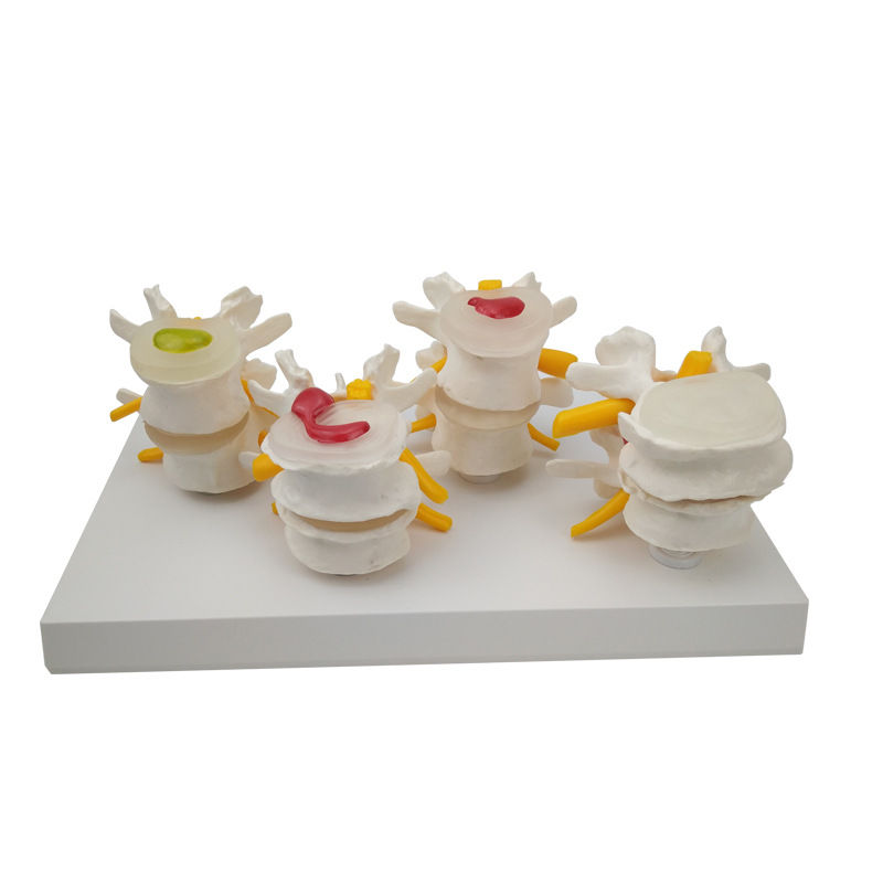

4 Phases Pathological Vertebra
This anatomical model is an advanced visual learning tool that simulates the pathological changes in the human lumbar intervertebral disc. It is designed to illustrate four distinct stages of degeneration — from a normal, healthy disc to varying degrees of deterioration, including herniation and other spinal pathologies. Each stage is meticulously crafted with precise anatomical detail, allowing for clear visualization of how intervertebral discs change over time due to mechanical stress and aging.
The model provides a side-by-side comparison between a healthy and a diseased lumbar spine, making it an indispensable tool for both theoretical and practical education. It helps explain the biomechanical aspects of spinal pathology and serves as a tangible reference for discussions on diagnosis, treatment, and rehabilitation. The flexible construction allows demonstration of dynamic disc movement, enhancing its pedagogical value.
This model is particularly suitable for use in medical classrooms, clinical demonstrations, and patient education. It enables students and healthcare professionals to examine detailed anatomical structures and serves as a powerful tool for individual study and comparison. Constructed from high-quality, durable PVC, it ensures long-lasting use, easy cleaning, and low maintenance.
Overall, this lumbar disc pathology model combines scientific accuracy with practical usability, offering a comprehensive understanding of the complex degenerative processes that occur in the lower spine.
Origin: Mainland China
Type: Skeletal Model
Subject: Medical Science
Material: PVC
Color: As shown
Size: 26.7 × 15.8 × 10 cm
Included: 1 × Model
Note: Minor color variations may occur due to lighting conditions. Product dimensions may vary by 0.5–1 cm due to manual measurement.


