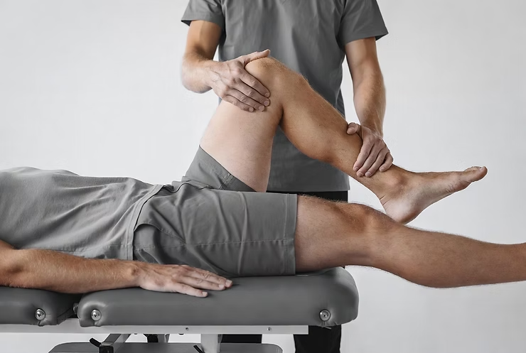
Gaenslen’s Test
- Fysiobasen

- Dec 18, 2025
- 5 min read
Gaenslen’s Test (Gaenslen’s Maneuver) is one of the five classical provocation tests used to identify musculoskeletal disorders and chronic inflammatory conditions of the lumbar spine and sacroiliac (SI) joint. The other commonly used SI joint pain provocation tests include the Distraction Test, Thigh Thrust Test, Compression Test, and Sacral Thrust Test.

When SI joint pain is suspected, evidence suggests that a cluster approach should be used – at least three positive tests – to improve diagnostic accuracy, particularly in patients with low back pain that does not centralize with repeated movement testing. Centralization is highly specific for discogenic pain, and in such cases, positive SI joint tests should be disregarded.
Gaenslen’s Test may also be helpful when evaluating pubic symphysis instability, hip joint pathology, L4 nerve root involvement, or femoral nerve stress. For this reason, it is often applied in the differential diagnosis of spondyloarthritis, sciatica, and other rheumatological conditions affecting the SI joint.
Purpose
The primary aims of Gaenslen’s Test are to:
Identify pain originating from the sacroiliac joint
Differentiate SI joint pathology from other causes of low back or pelvic pain
Support diagnostic certainty when combined with other SI joint provocation tests
Clinical Signs
Localized pain in the SI joint, hip joint, or pubic symphysis
Pain radiating along the L4 nerve root distribution
Reproduction of the patient’s familiar pain is considered a key positive indicator
Test Procedure
Patient position:
The patient lies supine with the painful leg positioned at the edge of the treatment table.
Asymptomatic leg:
The hip and knee are flexed to approximately 90°, and the patient holds the knee to the chest with both arms.
Symptomatic leg:
The examiner stabilizes the pelvis with one hand and applies gentle downward pressure with the other hand to extend the symptomatic leg beyond the edge of the table.
Movement:
This maneuver creates a torsional stress on the pelvis: the asymptomatic leg is forced into flexion while the symptomatic leg is extended.
This differential movement provokes the SI joint.
Tips:
In cases of bilateral pain, the test should be performed on both sides to assess asymmetry and pain localization.
Interpretation
Positive test: Reproduction of the patient’s typical pain in the SI joint, hip, or pubic symphysis. In some cases, it may also indicate L4 nerve root irritation.
Negative test: No pain or discomfort during the maneuver.
Evidence
Sensitivity: 37–61.5% (variable across studies)
Specificity: 33.3–100% (protocol-dependent)
Positive likelihood ratio (LR+): 1.02–2.29 (limited to moderate diagnostic utility)
Negative likelihood ratio (LR–): 0.65–1.11 (not sufficient to rule out SI pathology alone)
Inter-tester reliability (Kappa): 0.54–0.76
Best diagnostic accuracy is achieved when Gaenslen’s Test is positive in combination with at least two other SI joint provocation tests.
Clinical Relevance
Gaenslen’s Test is particularly valuable for:
Provoking pain from the sacroiliac joint in patients with non-centralizing low back pain
Differentiating SI joint dysfunction from hip joint pathology, pubic symphysis instability, or L4 radiculopathy
Assisting in the diagnostic work-up for spondyloarthritis, sciatica, and inflammatory spinal disorders
Following a positive test, further diagnostic confirmation with imaging (e.g., MRI) or diagnostic anesthetic injection into the SI joint is recommended. Fluoroscopic guidance is considered the gold standard for accurate needle placement.
Limitations
Low to moderate sensitivity and specificity – should not be used as a standalone test
Can yield false positives in cases of hip pathology, L4 root irritation, or pubic symphysis dysfunction
Must always be interpreted in the context of history, other clinical tests, and imaging findings
Summary
Gaenslen’s Test is a practical and widely used provocation test for assessing sacroiliac joint dysfunction. It should always be performed bilaterally and interpreted as part of a multi-test battery. A positive result is indicated by reproduction of the patient’s familiar pain in the SI joint or related pelvic structures. While easy to perform, the test has limited diagnostic accuracy in isolation and should be combined with other clinical assessments and, when necessary, advanced imaging for diagnostic confirmation.
Sources:
Gaenslen FJ. Sacro-iliac arthrodesis: indications, author's technic and end-results. Journal of the American Medical Association. 1927 Dec 10;89(24):2031-5.
Laslett M, Aprill CN, McDonald B, Young SB. Diagnosis of sacroiliac joint pain: validity of individual provocation tests and composites of tests. Manual therapy. 2005 Aug 1;10(3):207-18.
Laslett M. Pain provocation tests for diagnosis of sacroiliac joint pain. The Australian journal of physiotherapy. 2006;52(3):229.
↑ Jump up to:4.0 4.1 Dutton M. The shoulder complex. Dutton M. Orthopaedic Examination, Evaluation, and Intervention. 2nd ed. New York, NY: McGraw Hill Companies. 2008:523-4.
DDreyfuss P, Michaelsen M, Pauza K, McLarty J, Bogduk N. The value of medical history and physical examination in diagnosing sacroiliac joint pain. Spine. 1996 Nov 15;21(22):2594-602.
Cook C, Hegedus EJ. Orthopedic physical examination tests: An evidencebased approach. Upper Saddle River: Rearson Education.
Kokmeyer DJ, van der Wurff P, Aufdemkampe G, Fickenscher TC. The reliability of multitest regimens with sacroiliac pain provocation tests. Journal of Manipulative and Physiological Therapeutics. 2002 Jan 1;25(1):42-8.
Clinically Relevant Technologies, http://www.youtube.com/watch?v=Y2DrX6qy2yI; accessed May 2011
Flynn TW, Cleland J, Whitman J. Users’ guide to the musculoskeletal examination: fundamentals for the evidence-based clinician. Louisville, KY: Evidence in Motion. 2008.
Laslett M, Williams M. The reliability of selected pain provocation tests for sacroiliac joint pathology. Spine. 1994 Jun;19(11):1243-9.
Ozgocmen S, Bozgeyik Z, Kalcik M, Yildirim A. The value of sacroiliac pain provocation tests in early active sacroiliitis. Clinical rheumatology. 2008 Oct 1;27(10):1275-82.
Whiting P, Rutjes AW, Reitsma JB, Bossuyt PM, Kleijnen J. The development of QUADAS: a tool for the quality assessment of studies of diagnostic accuracy included in systematic reviews. BMC medical research methodology. 2003 Dec 1;3(1):25.
Whiting P, Harbord R, Kleijnen J. No role for quality scores in systematic reviews of diagnostic accuracy studies. BMC medical research methodology. 2005 Dec 1;5(1):19.
dde Graaf I, Prak A, Bierma-Zeinstra S, Thomas S, Peul W, Koes B. Diagnosis of lumbar spinal stenosis: a systematic review of the accuracy of diagnostic tests. Spine. 2006 May 1;31(10):1168-76.
Sehgal N, Shah RV, McKenzie-Brown AM, Everett CR. Diagnostic utility of facet (zygapophysial) joint injections in chronic spinal pain: a systematic review of evidence. Pain Physician. 2005 Apr;8(2):211-24.
Shah RV, Everett CR, McKenzie-Brown AM, Sehgal N. Discography as a diagnostic test for spinal pain: A systematic and narrative review. Pain Physician. 2005 Apr 1;8(2):187-209.
Hardaker Jr WT, Garrett Jr WE, Bassett 3rd FH. Evaluation of acute traumatic hemarthrosis of the knee joint. Southern medical journal. 1990 Jun 1;83(6):640-4.
Hegedus EJ, Cook C, Hasselblad V, Goode A, Mccrory DC. Physical examination tests for assessing a torn meniscus in the knee: a systematic review with meta-analysis. journal of orthopaedic & sports physical therapy. 2007 Sep;37(9):541-50.
McGrath MC. Clinical considerations of sacroiliac joint anatomy: a review of function, motion and pain. Journal of Osteopathic Medicine. 2004 Apr 1;7(1):16-24.


