Mast Cell Activation Syndrome (MCAS)
- Fysiobasen

- Dec 15, 2025
- 8 min read
Mast Cell Activation Syndrome (MCAS) is a little-known but significant condition affecting the immune system. The syndrome belongs to the group of mast cell disorders and is characterized by an overreaction of the body’s mast cells – cells that are normally activated to protect us against potentially harmful stimuli. In MCAS, mast cells respond inappropriately, often without any known trigger, and symptoms may vary greatly both between and within individuals. The condition is frequently mistaken for other diseases, which means it may remain undiagnosed for many years¹.

Immune Response Out of Control
Normally, mast cells are activated in response to allergens, heat, pressure, or infection. This can occur through either an IgE-mediated or non-IgE-mediated mechanism¹. The mast cells then release various pro-inflammatory substances such as histamine and prostaglandins to initiate a local immune reaction². In people with MCAS, this activation occurs excessively or inappropriately – without any other explanation for the reaction².
Three Types of Mast Cell Activation
There are three main forms of MCAS³:
Monoclonal (primary):– Caused by a genetic abnormality, often mutations in the KIT gene or aberrant expression of CD25/CD2– Associated with increased mast cell production, and also seen in systemic mastocytosis
Secondary (non-clonal):– Normal number of mast cells, but overactivation due to allergic reactions– May be IgE- or non-IgE-mediated
Idiopathic:– No known cause can be identified– Likely the most common form– Patients may show features of more than one type simultaneously¹
In severe cases, the condition can lead to systemic reactions and anaphylaxis.
Clinically Relevant Anatomy
Mast cells are granulated immune cells formed in the bone marrow and found in most tissues, particularly in the skin, connective tissue, intestines, and airways⁴. They are often located near blood vessels and the lymphatic system. Inside mast cells are mediators – such as histamine, leukotrienes, prostaglandins, tryptase, and cytokines – which are released upon activation⁵⁶.
For survival, mast cells must bind to stem cell factor (KIT), a transmembrane tyrosine kinase receptor⁷. Once they migrate to a tissue, they mature based on local signals such as interleukins (IL-3, 4, 9) and TGF-β1⁸⁹.
Two main mast cell types are distinguished:– MCT (tryptase): Found in connective tissue, skin, and peritoneum– MCTC (tryptase and chymase): Found in the intestines and airways⁸
Physiology and Pathology
Mast cell immune responses are triggered through different receptors: for IgE, estrogen, ATP, mechanical pressure, and toxins. Upon stimulation, mediators are synthesized and released, leading to increased inflammation, vasodilation, itching, pain, and in severe cases, circulatory collapse⁵⁶¹⁰.
In addition to their role in immune defense, mast cells are also involved in:– Tissue homeostasis– Phagocytosis– Cytokine production– Regulation of blood vessels and lymphatics¹¹
They reside within tissues, surrounded by blood vessels, nerves, and lymphatics – explaining why MCAS can cause such widespread and systemic symptoms.
Clinical Presentation
The symptom profile in MCAS is highly variable and depends on which mediators are released as well as which organ systems are affected. Reactions may change over time and vary with repeated exposure to the same stimulus. Many patients experience symptoms from the gastrointestinal tract.
Common Symptoms
Common symptoms include⁵¹²:
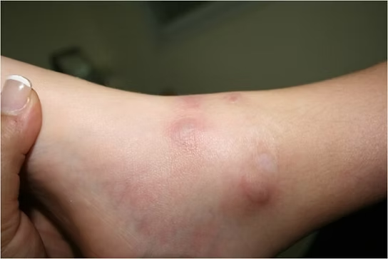
Skin: Flushing, itching, rashes, lesions
General: Fatigue, headaches, body pain
Skeletal: Osteoporosis, bone pain
Gastrointestinal: Bloating, abdominal pain, nausea, diarrhea, GERD
Respiratory: Throat swelling, shortness of breath
Circulatory: Cramps, hypotension, bleeding problems
Lymphatic: Enlarged lymph nodes, weight loss
Common Correlates and Overlapping Conditions
Frequently associated conditions include¹¹¹²¹³:
Ehlers–Danlos syndrome
Postural Orthostatic Tachycardia Syndrome (POTS)
Autoimmune diseases
Celiac disease and gluten intolerance
Fibromyalgia-like pain
Neurological and psychiatric symptoms
Chronic sinusitis and skin conditions
These correlations may raise suspicion but are not diagnostic criteria in themselves.
Diagnostic Criteria
Diagnosis of MCAS requires the following three criteria¹¹¹³:
Systemic symptoms involving at least two organ systems
Detectable mast cell activation (e.g., elevated tryptase)
Response to mast cell–targeted treatment
Tests Used
Serum tryptase: Sample taken within 4 hours after reaction and compared to baseline. Interpretation requires that levels exceed baseline by 20% + 2 ng/ml.
Urine tests: For mediators such as N-methylhistamine, prostaglandin D2, leukotriene E4.
Treatment response: Symptom relief from antihistamines or mast cell stabilizers supports the diagnosis.
Endoscopy: May show mast cells in the intestine but rarely used routinely.
Treatment and the Role of Physiotherapy
Treatment is aimed at reducing symptoms and limiting mediator release:
Antihistamines (H1- and H2-blockers)
Mast cell stabilizers (cromolyn)
Leukotriene antagonists
Corticosteroids for severe episodes
Elimination diet when food reactions are suspected
Reduction of psychosocial stress
Physiotherapy’s Role
Physiotherapists play an important role, especially in the following areas:
Stress management and breathing: MCAS patients may develop dysfunctional breathing patterns (e.g., hyperventilation) that worsen symptoms. Breathing techniques and relaxation can reduce reactions.
Adapted physical activity: Regular exercise may help stabilize immune responses and support overall health.
Awareness of hypersensitivity: When treating patients with EDS, POTS, or autoimmune conditions, physiotherapists should keep MCAS in mind as a differential diagnosis.
It is also important to understand the link between trauma and mast cell disorders – particularly in women’s health. Physiotherapists can act as bridges to other services and provide reassurance throughout the patient journey.
Challenges in Diagnosis
Diagnosing Mast Cell Activation Syndrome (MCAS) is difficult due to the highly variable symptom profile. To establish the diagnosis, three criteria must be met¹¹¹³:
Systemic symptoms involving at least two organ systems
Objective evidence of mast cell activation (biochemical markers or cell increase)
Clinical response to mast cell–targeted treatment
Organs Commonly Involved in MCAS
Ear, nose, and throat
Gastrointestinal tract
Cardiovascular system: dizziness, inflammation, unstable heart rate
Skeletal system: bone pain, osteoporosis
Skin: rashes, flushing
Urinary tract: bladder irritation
Tests and Measurements

Serum tryptase: Gold standard. The blood sample must be taken within 4 hours of symptom onset, and levels must be >20% above baseline + 2 ng/ml. If no baseline is available, a new sample must be taken 24–48 hours after the episode¹¹.
Urine tests: Should be analyzed within 24 hours for mediators such as N-methylhistamine, prostaglandin D2, F2α, and leukotriene E4⁵.
Treatment response: If symptoms are reduced after treatment with mast cell stabilizers, this supports the diagnosis – even when objective findings are lacking.
Endoscopy: May detect mast cells in the GI tract but is rarely used diagnostically due to its invasiveness.
Treatment and Management
Treatment consists of both medication and lifestyle measures. Identifying and avoiding triggers is essential – but often difficult, as reactions can be unpredictable. Stress is a well-known aggravating factor and should be addressed systematically¹³.
In IgE-related MCAS, immunotherapy may be considered¹⁴–¹⁶.
Pharmacological Treatment
H1-antihistamines: Relieve acute symptoms such as itching, rashes, and headaches
1st generation: hydroxyzine, diphenhydramine, doxepin, ketotifen
2nd generation: cetirizine, fexofenadine, loratadine
H2-antihistamines: Stabilize mast cells and relieve GI symptoms
famotidine, cimetidine
Mast cell stabilizers and glucocorticoids: Reduce symptoms such as brain fog and GI issues
Tyrosine kinase inhibitors: Broad effect on mast cells and IgE response
High-dose aspirin: For elevated prostaglandins (brain fog, flushing, bone pain)¹⁷–¹⁹
Treatment must be individually tailored and often combined to achieve effect¹⁸.
Differential Diagnoses
Because MCAS resembles many other conditions, thorough differential diagnostics are required¹³. Relevant conditions include:
Infections
IBS and celiac disease
Adrenal insufficiency
Cardiovascular disorders
Psychiatric and neurological conditions
Endocrine diseases
Toxic reactions
Other mast cell diseases that must be excluded:
Systemic mastocytosis (SM)
Cutaneous mastocytosis (CM)
Smoldering systemic mastocytosis (SSM)
Hereditary alpha-tryptasemia (HaT)
Relevance for Physiotherapy
MCAS patients often have many everyday triggers – several of which may occur in clinical practice. Physiotherapists should therefore be aware of factors such as:
Physical activity (exertion)
Temperature changes
Friction, vibration, and pressure
Medications (opioids, NSAIDs, local anesthetics)²⁰–²²
Patients should also be protected from known allergens, food intolerances, emotional stress, and infections.
MCAS, Connective Tissue and Autoimmunity
Mast cells are present in connective tissue and are believed to play a role in conditions such as Ehlers–Danlos syndrome (EDS) and Hypermobility Spectrum Disorders (HSD). It has been hypothesized that mast cell mediators – such as tryptase and histamine – affect fibroblast proliferation and collagen synthesis²³²⁴.
Several clinical similarities exist between MCAS and hEDS/HSD, and recognizing this relationship may help physiotherapists identify undiagnosed patients²³.
Urticaria and Autoimmune Conditions
Chronic urticaria is frequently observed in both EDS and MCAS and is likely caused by mast cell activity²³²⁵²⁶. Overlap between MCAS and lupus (SLE) has also been reported in patients with persistent urticaria²⁶.
Rheumatoid Arthritis and Mast Cells
Mast cells are found in increased numbers in the joints of people with rheumatoid arthritis (RA) and contribute to the disease process²⁷²⁸. Different synovitis phenotypes have been identified based on mast cell density, and IgE-mediated activity may contribute to joint destruction²⁹. This provides an important pathophysiological link between MCAS and rheumatic diseases.ykdommer.
References
Akin C. Mast cell activation syndromes. J Allergy Clin Immunol. 2017 Aug;140(2):349–355. doi:10.1016/j.jaci.2017.06.007. PMID: 28780942.
Anvari S, Miller J, Yeh CY, Davis CM. IgE-Mediated Food Allergy. Clin Rev Allergy Immunol. 2019 Oct;57(2):244–260. doi:10.1007/s12016-018-8710-3. PMID: 30370459.
The Mast Cell Disease Society. Mast Cell Activation Syndrome Variants. Available from: https://tmsforacure.org/overview/mast-cell-activation-syndrome-variants/ (last accessed 05.07.2025).
Moon TC, St Laurent CD, Morris KE, Marcet C, Yoshimura T, Sekar Y, Befus AD. Advances in mast cell biology: new understanding of heterogeneity and function. Mucosal Immunol. 2010 Mar;3(2):111–128. doi:10.1038/mi.2009.136. PMID: 20043008.
Valent P, Akin C, Arock M, Brockow K, Butterfield JH, Carter MC, et al. Definitions, criteria and global classification of mast cell disorders with special reference to mast cell activation syndromes: a consensus proposal. Int Arch Allergy Immunol. 2012;157(3):215–225.
Valent P. Mast cell activation syndromes: definition and classification. Allergy. 2013 Apr;68(4):417–424.
Fong M, Crane JS. Histology, Mast Cells. [Updated May 8, 2022]. In: StatPearls [Internet]. Treasure Island (FL): StatPearls Publishing. Available from: https://www.ncbi.nlm.nih.gov/books/NBK499904/
Galli SJ, Borregaard N, Wynn TA. Phenotypic and functional plasticity of cells of innate immunity: macrophages, mast cells and neutrophils. Nat Immunol. 2011 Oct;12(11):1035–1044. doi:10.1038/ni.2109. PMID: 22012443.
Cardamone C, Parente R, Feo GD, Triggiani M. Mast cells as effector cells of innate immunity and regulators of adaptive immunity. Immunol Lett. 2016 Oct;178:10–14. doi:10.1016/j.imlet.2016.07.003. PMID: 27393494.
Valent P, Akin C, Bonadonna P, Hartmann K, Brockow K, Niedoszytko M, et al. Proposed Diagnostic Algorithm for Patients with Suspected Mast Cell Activation Syndrome. J Allergy Clin Immunol Pract. 2019 Apr;7(4):1125–1133.e1. doi:10.1016/j.jaip.2019.01.006.
Theoharides TC, Valent P, Akin C. Mast cells, mastocytosis, and related disorders. N Engl J Med. 2015 Jul 9;373(2):163–172.
Matito A, Escribese MM, Longo N, Mayorga C, Luengo-Sánchez O, et al. Clinical Approach to Mast Cell Activation Syndrome: A Practical Overview. J Investig Allergol Clin Immunol. 2021 Dec 21;31(6):461–470. doi:10.18176/jiaci.0675. PMID: 33541851.
Niedoszytko M, Bonadonna P, Elberink JNGO, Golden DBK. Epidemiology, diagnosis, and treatment of hymenoptera venom allergy in mastocytosis patients. Immunol Allergy Clin North Am. 2014;34:365–381. doi:10.1016/j.iac.2014.02.004.
Bonadonna P, Bonifacio M, Lombardo C, Zanotti R. Hymenoptera allergy and mast cell activation syndromes. Curr Allergy Asthma Rep. 2016;16. doi:10.1007/s11882-015-0582-5.
Valent P, Akin C, Nedoszytko B, Bonadonna P, Hartmann K, et al. Diagnosis, Classification and Management of Mast Cell Activation Syndromes (MCAS) in the Era of Personalized Medicine. Int J Mol Sci. 2020 Nov 27;21(23):9030. doi:10.3390/ijms21239030.
Valent P, Akin C, Hartmann K, George TI, Sotlar K, et al. Midostaurin: A magic bullet that blocks mast cell expansion and activation. Ann Oncol. 2017;28:[[1]]. doi:10.1093/annonc/mdx290.
Krauth MT, Mirkina I, Herrmann H, Baumgartner C, Kneidinger M, Valent P. Midostaurin (PKC412) inhibits immunoglobulin E-dependent activation and mediator release in human blood basophils and mast cells. Clin Exp Allergy. 2009;39:1711–1720. doi:10.1111/j.1365-2222.2009.03353.x.
Boyden SE, Desai A, Cruse G, Young ML, Bolan HC, Scott LM, et al. Vibratory Urticaria Associated with a Missense Variant in ADGRE2. N Engl J Med. 2016 Feb 18;374(7):656–663.
Akin C, Metcalfe DD. Mastocytosis and mast cell activation syndromes presenting as anaphylaxis. In: Castells MC, ed. Anaphylaxis and Hypersensitivity Reactions. New York: Humana Press; 2011. p. 245–256. doi:10.1007/978-1-60327-951-2_15.
Jennings S, Russell N, Jennings B, Slee V, Sterling L, Castells M, et al. The Mastocytosis Society survey on mast cell disorders: patient experiences and perceptions. J Allergy Clin Immunol Pract. 2014 Jan–Feb;2(1):70–76.
Monaco A, Choi D, Uzun S, Maitland A, Riley B. Association of mast-cell-related conditions with hypermobile syndromes: a review of the literature. Immunol Res. 2022 Aug;70(4):419–431. doi:10.1007/s12026-022-09280-1. PMID: 35449490.
Seneviratne SL, Maitland A, Afrin L. Mast cell disorders in Ehlers-Danlos syndrome. Am J Med Genet C Semin Med Genet. 2017;175:226–236. doi:10.1002/ajmg.c.31555.
Szalewski RJ, Davis BP. Ehlers-Danlos Syndrome is associated with Idiopathic Urticaria – a retrospective study [abstract]. J Allergy Clin Immunol. 2019;143. doi:10.1016/j.jaci.2018.12.204.
Greiwe J. An index case of a rare form of inducible urticaria successfully treated with omalizumab [abstract]. Ann Allergy Asthma Immunol. 2018;121:S84. doi:10.1016/j.anai.2018.09.273.
Pitzalis C, Kelly S, Humby F. New learnings on the pathophysiology of RA from synovial biopsies. Curr Opin Rheumatol. 2013;25:334–344.
Malmstrom V, Catrina AI, Klareskog L. The immunopathogenesis of seropositive rheumatoid arthritis: from triggering to targeting. Nat Rev Immunol. 2017;17:60–75.
Crisp AJ, Chapman CM, Kirkham SE, Schiller AL, Krane SM. Articular mastocytosis in rheumatoid arthritis. Arthritis Rheum. 1984;27:845–851.
Conti P, Kempuraj D. Important role of mast cells in multiple sclerosis. Mult Scler Relat Disord. 2016 Jan;5:77–80. doi:10.1016/j.msard.2015.11.005


