Meningitis
- Fysiobasen

- Dec 24, 2025
- 6 min read
Meningitis is an infectious disease of the central nervous system that causes inflammation of the three meningeal layers (dura mater, arachnoid, and pia mater) surrounding the brain and spinal cord.[1] Before the antibiotic era the disease was uniformly fatal, and even today meningitis carries a mortality of up to 25%.[2] Multiple pathogens can cause meningitis—including bacteria, fungi, and viruses—but the greatest global burden is from bacterial meningitis.[3] Even with modern diagnostics, therapy, and vaccination, there were an estimated 8.7 million cases of meningitis worldwide in 2015 with 379,000 deaths. The first reported case of COVID-19–associated meningitis appeared early in 2020, and some reports warn that SARS-CoV-2 may have neuroinvasive potential because certain patients present with meningitic symptoms (e.g., headache, nausea, vomiting).[4]

Causes
Meningitis is defined as inflammation of the meninges. The meninges are the three layers (dura mater, arachnoid, pia mater) that envelop the brain and spinal cord (whereas encephalitis is inflammation of the brain parenchyma itself).
Meningitis may result from infectious or non-infectious processes (autoimmune diseases, cancer/paraneoplastic syndromes, drug reactions).
Infectious causes include bacteria, viruses, fungi, and more rarely parasites.[5]
Risk Factors
Chronic illnesses (renal failure, diabetes, cystic fibrosis)
Extremes of age
Incomplete vaccination
Immunodeficiency (iatrogenic, transplants, AIDS)
Crowded living conditions
Travel to endemic regions
Alcohol use
Head trauma or neurosurgery
Intravenous drug use
Sickle-cell disease and prior splenectomy
Infections of the sinuses, ear, or mastoid[1][2]
Meningococcal Meningitis
This entity is especially important because it can cause large epidemics. The bacterium spreads via respiratory droplets from carriers. All ages can be affected, but infants, preschool children, and adolescents are at highest risk. Untreated meningococcal meningitis is fatal in up to 50% of cases; among survivors, 10–20% suffer brain damage, hearing loss, or other disabilities.[6] Although vaccines have existed for >40 years, there is still no universal vaccine that covers all strains.[7]
Epidemiology
Incidence in high-income countries is 2–6 per 100,000 adults per year, but up to 10× higher in low-income settings.[8][9] In the United States, bacterial meningitis occurs at roughly 1.38 cases per 100,000 with a 14.3% mortality. Globally, the highest numbers occur in Africa’s “meningitis belt,” from Ethiopia to Senegal. Incidence has fallen substantially over the last 15 years thanks to vaccination programs.[1]
Common pathogens:
Bacteria: Streptococcus pneumoniae, group B streptococci, Neisseria meningitidis, Haemophilus influenzae, Listeria monocytogenes[10]
Viruses: Non-polio enteroviruses (group B coxsackieviruses, echoviruses); herpesviruses (EBV, HSV, VZV); mumps, measles; arboviruses (West Nile, La Crosse, Powassan)
Fungi: Cryptococcus neoformans, Coccidioides immitis, Aspergillus, Candida, Mucorales—especially in diabetes or transplant recipients[2][11]
Pathophysiology
Meningitis typically develops via two routes:
Hematogenous spread: Bacteria colonize the nasopharynx, invade the bloodstream, cross the blood–brain barrier, and trigger meningeal inflammation.
Direct spread: From adjacent structures (ear, sinuses), foreign bodies, or surgery. Viruses may also reach the CNS via retrograde axonal transport or hematogenous
spread.[2]
Symptoms & Clinical Presentation
Headache, fever, vomiting, and neck stiffness are the most common symptoms.[1][12][13]
Early features include nausea, drowsiness, and confusion.
Pain in the thighs or low back may occur.[1]
Later signs include seizures, photophobia, and tachypnea.
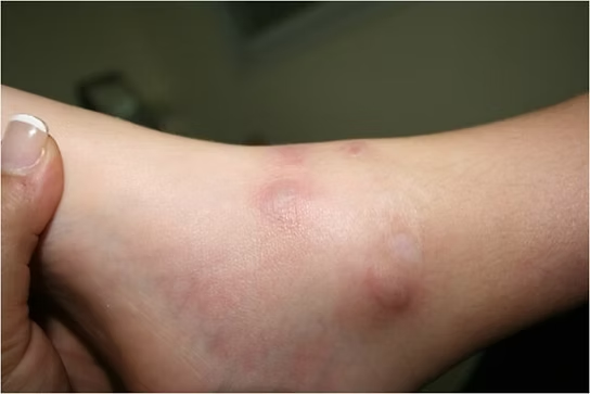
Rash (petechiae or purpura) occurs in 80–90% of bacterial cases.[13][14][15]
Meningeal inflammation can provoke pain with passive neck flexion (Brudzinski’s sign) or with passive knee extension (Kernig’s sign).[16] Untreated bacterial meningitis may involve the brain parenchyma, causing lethargy, vomiting, seizures, papilledema, confusion, coma, focal neurologic deficits, and cranial neuropathies.
Treatment & Management
Antibiotics plus supportive care are critical in all bacterial meningitis.
Airway management, oxygenation, adequate IV fluids, and fever control are foundational.
Antibiotic therapyChoice depends on the likely pathogen; clinicians should consider patient demographics and history to ensure optimal empiric coverage.
Steroid therapyEvidence is insufficient to recommend widespread steroid use across all bacterial meningitis cases.
Raised Intracranial Pressure (ICP)If clinical signs of elevated ICP develop (altered mental status, neurologic deficits, non-reactive pupils, bradycardia), measures to maintain cerebral perfusion may include:
Head of bed elevated to 30°
Mild hyperventilation in intubated patients
Osmotic therapy such as 25% mannitol or 3% hypertonic saline
ChemoprophylaxisIndicated for close contacts of patients with N. meningitidis and H. influenzae type b meningitis. This includes household members, partners, utensil-sharers, and healthcare workers with close exposure to secretions (mouth-to-mouth resuscitation, unmasked intubation).[2]
Diagnostic Tests
Diagnosis relies on cerebrospinal fluid (CSF) analysis—WBC count, glucose, protein, culture, and, in selected cases, PCR. CSF is obtained via lumbar puncture, and opening pressure can be measured.
Additional testing by suspected etiology:
Viruses: Multiplex PCR and pathogen-specific PCRs
Fungi: CSF fungal culture, India ink for Cryptococcus
Tuberculosis: CSF with Ziehl–Neelsen staining and culture
Syphilis: CSF VDRL
Borrelia: CSF Borrelia antibody[2]
Complications
A 2010 meta-analysis reported a median 19.9% risk of sequelae after discharge. Most commonly isolated organisms were H. influenzae and S. pneumoniae. Frequent sequelae included:
Hearing loss (6%)
Behavioral problems (2.6%)
Cognitive difficulties (2.2%)
Motor deficits (2.3%)
Epilepsy (1.6%)
Visual problems (0.9%)
Other complications:
Elevated ICP from cerebral edema (due to BBB disruption, cytotoxic cytokines, immune response, and bacteria)
Hydrocephalus
Cerebrovascular complications
Focal neurologic deficits[2][12][13]
Physiotherapy Management
Per the American Physical Therapy Association’s Guide to Physical Therapist Practice, CNS infections align with:
5D: Impaired motor function and sensory integrity associated with non-progressive CNS disorders acquired in adulthood
5I: Impaired arousal, ROM, and motor control associated with coma, near coma, or vegetative state
Physiotherapy often begins in the ICU. When planning care, review the chart and note contraindications such as ICP, cerebral perfusion pressure, and other key labs. Meningitis may produce deficits similar to brain injury, neurologic complications, immune dysfunction, and secondary functional limitations.
Understanding consciousness level and behavioral changes helps tailor treatment. Modify the environment to reduce light/sound hypersensitivity, and monitor vital signs to gauge response. Familiarity with the Glasgow Coma Scale and frequent reassessment are important.
Early positioning and range-of-motion work in the acute phase are essential. Proper positioning with pillows/towels helps prevent pressure injuries and contractures. Maintaining cervical and trunk mobility supports functional movement. Early mobilization lowers the risk of secondary complications and improves prognosis.
Education for the patient, family, and caregivers is crucial: condition overview, illness stages, secondary complications, red flags, and resources foster engagement. Emphasize potential systemic involvement and that rehabilitation may take time and vary by individual.[1]
Differential Diagnosis
Stroke
Subdural hematoma
Subarachnoid hemorrhage
Brain metastases
Brain abscess (can co-exist with meningitis)[2]
References
Goodman C, Fuller K. Pathology: Implications for the Physical Therapist. 3rd ed. St. Louis, Missouri: Saunders Elsevier, 2009.
Hersi K, Kondamudi NP. 2020 Meningitis.:https://www.ncbi.nlm.nih.gov/books/NBK459360/
WHO Meningitis :https://www.who.int/health-topics/meningitis#tab=tab_1
Moriguchi T, Harii N, Goto J, Harada D, Sugawara H, Takamino J, Ueno M, Sakata H, Kondo K, Myose N, Nakao A. A first case of meningitis/encephalitis associated with SARS-Coronavirus-2. International Journal of Infectious Diseases. 2020 Apr 3.:https://www.sciencedirect.com/science/article/pii/S1201971220301958
Hersi K, Gonzalez FJ, Kondamudi NP. Meningitis. In: StatPearls [Internet]. Treasure Island (FL): StatPearls Publishing; 2021 Jan-. : https://www.ncbi.nlm.nih.gov/books/NBK459360/
World Health Organization. Meningococcal meningitis. : https://www.who.int/news-room/fact-sheets/detail/meningococcal-meningitis
WHO Meningitis :https://www.who.int/westernpacific/health-topics/meningitis
Goodman CC, Fuller KS. Pathology: implications for the physical therapist. 4th ed. St. Louis, MO: Elsevier Saunders; 2015.
Meningitis Belt [Internet]. 2017 https://wwwnc.cdc.gov/travel/yellowbook/2016/infectious-diseases-related-to-travel/meningococcal-disease
gjennomgått -Trukket
Meningococcal Graph [Internet]. 2017: https://www.cdc.gov/meningococcal/surveillance/
Porter RS, Kaplan JL. The Merck manual of diagnosis and therapy. Merck Sharp & Dohme Corp.; 2011.
Aminoff M, Greenberg D, Simon R. Clinical Neurology. 6th ed. New York, NY: Lange Medical Books/McGraw-Hill, 2005.
Paul N, Bowe C, Morrow G. Bacterial Meningitis. WIN. 2016;24(8):47-49.
Watkins J. Recognising the signs and symptoms of meningitis. British Journal of School Nursing. 2012;7(10):481-483.
Meningitis control in countries of the African meningitis belt, 2015. World Health Organization [Internet]. 2015;16:209-216. :http://eds.b.ebscohost.com.libproxy.bellarmine.edu/ehost/pdfviewer/pdfviewer?sid=f38a50a1-dce1-4c95-8af1-4b603ccaee02%40sessionmgr102&vid=9&hid=104
Neisseri Meningitidis. Brudzinski’s sign. http://bioweb.uwlax.edu/bio203/s2008/bingen_sama/neck.jpg
National Library of Medicine. Kernig’s sign. http://www.nlm.nih.gov/medlineplus/ency/images/ency/fullsize/19077.jpg
Gjennomgått - Trukket
Goodman CC, Fuller KS. Pathology: implications for the physical therapist. 4th ed. St. Louis, MO: Elsevier Saunders; 2015.
Valcour V, Haman A, Cornes S, Lawall C, Parsa A, Glaser C, et al. A case of enteroviral meningoencephalitis presenting as rapidly progressive dementia. Nature Clinical Practice. Neurology [serial on the Internet]. (2008, July)4(7): 399-403. : MEDLINE.
Thompson H. Not your "typical patient": cryptococcal meningitis in an immunocompetent patient. Journal of Neuroscience Nursing [serial on the Internet]. (2005, June), 37(3): 144-148. : CINAHL with Full Text.
Samuels M, Gonzalez R, Kim A, Stemmer-Rachamimov A. Case records of the Massachusetts General Hospital. Case 34-2007. A 77-year-old man with ear pain, difficulty speaking, and altered mental status. The New England Journal Of Medicine [serial on the Internet]. (2007, Nov 8), 357(19): 1957-1965. : MEDLINE.


