Scleroderma (Systemic Sclerosis)
- Fysiobasen

- Dec 24, 2025
- 5 min read
Scleroderma is a rare connective tissue disease of unknown pathogenesis, characterized by autoimmune inflammation and excessive collagen deposition in the skin, blood vessels, and internal organs. This leads to a chronic, progressive condition with wide variability in course and organ involvement. Progressive fibrosis thickens skin and soft tissues and can gradually impair organ function.¹–³

Classification
Scleroderma is broadly divided into systemic and localized forms.³
Systemic scleroderma (SSc) involves the skin and multiple organ systems (heart, lungs, kidneys, gastrointestinal tract, and musculoskeletal system).⁴ Subtypes:
Diffuse cutaneous SSc: Rapidly progressive skin thickening over large body areas with higher risk of internal organ fibrosis.⁵
Sine scleroderma: Internal organ fibrosis without cutaneous involvement; rare and diagnostically challenging.⁶
Limited cutaneous SSc (CREST): Skin involvement limited to fingers, hands, and face with slow progression. CREST = Calcinosis, Raynaud’s phenomenon, Esophageal dysmotility, Sclerodactyly, Telangiectasia.⁵
Localized scleroderma typically affects only skin and subcutaneous tissues in circumscribed areas.³
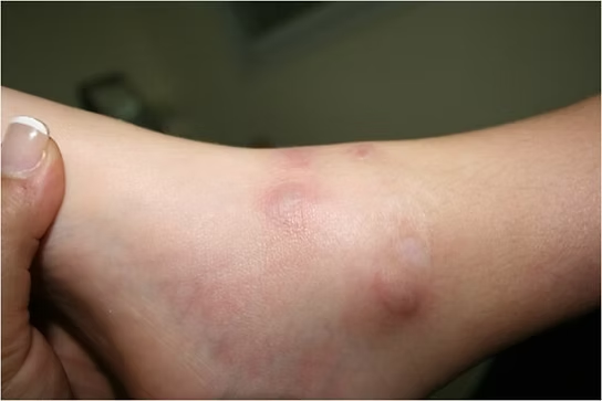
Morphea: Waxy, indurated plaques of variable size, shape, and color; may resolve over 3–5 years, though some develop deeper tissue involvement.
Linear scleroderma: A linear, band-like lesion on an arm, leg, scalp, or neck that often involves deeper tissues and restricts joint motion.
Overlap/Mixed Connective Tissue Disease (MCTD): Scleroderma features occurring with other systemic rheumatic diseases (e.g., SLE or polymyositis).⁷
Epidemiology
Scleroderma is uncommon, with prevalence varying by ethnicity, sex, and geography.⁸
SSc is more prevalent in Europe and the Americas than in East Asia, and highest among some Indigenous groups in Canada (~47/100,000).⁹
Women are affected ~4.6× more often than men, but men frequently experience more severe disease.⁹
~30% present with systemic disease; ~10% with sine scleroderma.⁹
Localized scleroderma is more common in children than adults.³
U.S. estimates: ~75,000–300,000 affected; peak onset 20–50 years.¹⁰
Genetic background and ethnicity influence phenotype and organ involvement (e.g., greater severity reported in Choctaw Native Americans and African Americans compared with those of European ancestry).¹¹
Diagnostic Workup (Key Tests & Labs)
Diagnosis can be difficult due to overlap with other conditions.¹² It relies on history, examination, and targeted testing:
Imaging:
X-ray/CT for skeletal changes.
MRI/ultrasound for soft-tissue assessment.¹²
Serology (selected):
Anti–topoisomerase I (Scl-70): linked to diffuse SSc.
Anticentromere antibodies (ACA): associated with CREST.
Elevated ESR: active disease/flare.
Anti-U1 RNP: overlap syndromes.¹² ¹³
Skin biopsy: Increased collagen deposition and adnexal atrophy in involved skin confirms cutaneous fibrosis.¹³
Not all patients have disease-specific antibodies, and seropositivity does not always equal clinical disease.¹³Early/rapid skin changes make diagnosis more straightforward; gradual courses may require months to years and exclusion of mimics.¹³ ¹⁴
Etiology (Proposed Mechanisms)
The precise cause remains unknown and non-infectious.¹⁴ Contributing factors likely include:
Immune dysregulation: Aberrant activation drives fibroblasts to overproduce collagen (fibrosis). Co-existing autoimmune disease increases risk.¹⁵
Genetics: Polygenic susceptibility (e.g., HLA, IRF5, STAT4 variants).¹⁵
Environment: Infections and certain chemicals may trigger disease in genetically susceptible individuals.¹⁴ ¹⁵
Hormonal influences: Female predominance suggests a role for estrogen and related pathways (evidence remains incomplete).¹¹ ¹⁴
Multisystem Involvement
Organ Manifestations at a Glance
System | Common Manifestations |
Skin | Swollen fingers/hands; sclerodactyly (tight, thick skin limiting mobility); telangiectasias |
Gastrointestinal | GERD; diarrhea from dysmotility/SIBO and malabsorption; constipation from smooth muscle fibrosis |
Lungs | Interstitial lung disease (fibrosis) causing impaired gas exchange; pulmonary arterial hypertension (PAH); reduced ventilatory function |
Heart | Heart failure (often secondary to PAH); pericarditis; arrhythmias |
Vascular | Digital ulcers; Raynaud’s phenomenon (cold/stress-induced color change of digits) |
Endocrine/Exocrine | Sicca features/Sjögren’s (dry eyes/mouth) |
Genitourinary | Hypertension, proteinuria; renal crisis (malignant hypertension, acute kidney failure); sexual dysfunction (ED, reduced lubrication) |
Dental/Oral | Caries (low saliva, GERD acidity); microstomia and limited oral aperture |
Medical Management
No current therapy halts or reverses collagen overproduction.¹ Treatment aims to control symptoms, limit complications, and preserve organ function, tailored to individual needs. Multidisciplinary care (often led by a rheumatologist) is standard, with dermatology, nephrology, cardiology, gastroenterology, pulmonology, and others involved as indicated.¹⁴
Pharmacologic Options (selected)¹⁰ ¹⁶
Inflammation/pain: NSAIDs; short courses of corticosteroids (use cautiously).
Immunosuppression (organ-threatening disease): Methotrexate, mycophenolate, cyclophosphamide (agent depends on target organ and risk profile).
Organ-targeted therapies:
GERD: Proton pump inhibitors.
Renal/HTN: ACE inhibitors (first-line for scleroderma renal crisis).
Skin: Phototherapy in selected cases.
ILD: Mycophenolate or cyclophosphamide.
PAH: Prostacyclin analogues, endothelin receptor antagonists, PDE-5 inhibitors.
Raynaud’s/ulcers: Calcium-channel blockers; PDE-5 inhibitors; topical/IV prostanoids (specialist care).
Sjögren’s features: Sialogogues, local measures.
Physiotherapy & Rehabilitation
Physiotherapy helps prevent contractures, maintain motion, manage pain/stiffness, and support daily function—often in partnership with occupational therapy.⁸ ¹⁴ ³ Programs are individualized to:
Reduce pain.
Improve strength.
Preserve/improve range of motion (ROM).
Prevent joint contractures.
Enhance circulation and protect fragile skin.
Promote independence in ADLs.
Modalities & Methods: Massage, hydrotherapy, neuromuscular electrical stimulation, ROM and joint mobilization.¹⁷ Evidence from controlled trials is mixed—tailor to phenotype, organ status, and tolerance.¹⁸
Exercise types:
Aerobic training: Walking, cycling, dancing to improve cardiopulmonary fitness and circulation.¹⁸
Strength training: Progressive resistance or functional strengthening (e.g., sit-to-stand, carrying bags).
Flexibility/mobility: Daily stretching, gentle yoga to counter stiffness and microstomia-related limitations (facial/oral exercises as needed).
Group or home-based formats can be effective when monitored and adapted for Raynaud’s, skin fragility, and cardiopulmonary limitations.
Include psychology/social work for coping and adherence; dental, orthodontic, and speech therapy for oral complications when needed.¹⁴ ³
Prognosis
Scleroderma is serious and associated with increased mortality, though outcomes have improved markedly; 5-year survival now approaches ~80%.¹ PAH portends worse outcomes (≈50% 2-year survival).¹ Patients with SSc-PAH generally fare worse than those with idiopathic PAH, underscoring the need for early detection and targeted therapy.
Differential Diagnosis (Selected)
Conditions that can mimic aspects of scleroderma include:¹⁹
Eosinophilic fasciitis (EF): Fascial involvement, typically spares fingers.
Nephrogenic systemic fibrosis: Gadolinium-associated fibrosis in renal impairment.
Scleroderma-like skin thickening: Scleromyxedema, graft-versus-host disease, porphyria cutanea tarda, “human adjuvant disease.”
Isolated Raynaud’s phenomenon.
A careful clinical evaluation with biopsy and relevant labs is essential to distinguish these entities.
References
Adigun R, Goyal A, Hariz A. Systemic sclerosis. :https://www.ncbi.nlm.nih.gov/books/NBK430875/
Rosendahl AH, Schönborn K, Krieg T. Pathophysiology of systemic sclerosis (scleroderma). The Kaohsiung journal of medical sciences. 2022 Mar;38(3):187-95.
Rosendahl AH, Schönborn K, Krieg T. Pathophysiology of systemic sclerosis (scleroderma). The Kaohsiung journal of medical sciences. 2022 Mar;38(3):187-95.
Arthritis Foundation. Scleroderma information sheet. https://www.arthritis.org/about-arthritis/types/scleroderma
Careta MF, Romiti R. Localized scleroderma: clinical spectrum and therapeutic update. Anais brasileiros de dermatologia. 2015 Jan;90:62-73.
Sato F, Sato M, Yamano T, Yamaguchi K, Miyake T. A Case of Mixed Connective Tissue Disease That Transformed Into Systemic Lupus Erythematosus After a Long Clinical Course. Cureus. 2023 Apr 27;15(4).
Calderon LM, Pope JE. Scleroderma epidemiology update. Current Opinion in Rheumatology. 2021 Mar 1;33(2):122-7.
Odonwodo A, Badri T, Hariz A. Scleroderma. InStatPearls [Internet] 2022 Aug 1. StatPearls Publishing.:https://www.ncbi.nlm.nih.gov/books/NBK537335/
Lescoat A, Huang S, Carreira PE, Siegert E, de Vries-Bouwstra J, Distler JH, Smith V, Del Galdo F, Anic B, Damjanov N, Rednic S. Cutaneous Manifestations, Clinical Characteristics, and Prognosis of Patients With Systemic Sclerosis Sine Scleroderma: Data From the International EUSTAR Database. JAMA dermatology. 2023 Aug 1;159(8):837-47. BibTeXEndNoteRefManRefWorks
American Academy of Dermatology Association. scleroderma: diagnosis and treatment.
Mayo Foundation for Medical Education and Research. Patient Care and Health Information. Sclerodermahttp://www.mayoclinic.com/health/scleroderma/DS00362
Marzano AV, Menni S, Parodi A, Borghi A, Fuligni A, Fabbri P, Caputo R. Localized scleroderma in adults and children. Clinical and laboratory investigations on 239 cases. European Journal of Dermatology. 2003 Apr 15;13(2):171-6.
Careta MF, Romiti R. Localized scleroderma: clinical spectrum and therapeutic update. Anais brasileiros de dermatologia. 2015 Jan;90:62-73.
National Institute of Arthritis and Musculoskeletal and Skin Diseases. Health Topics. Scleroderma.
US National Library of Medicine. Genetics Home Reference. Systemic Scleroderma. http://ghr.nlm.nih.gov/condition/systemicscleroderma /
Shah AA, Wigley FM. My approach to the treatment of scleroderma. In Mayo Clinic Proceedings 2013 Apr 1 (Vol. 88, No. 4, pp. 377-393).
Poole JL. Musculoskeletal rehabilitation in the person with scleroderma. Current opinion in rheumatology. 2010 Mar 1;22(2):205-12.
Frade S, Cameron M, Espinosa-Cuervo G, Suarez-Almazor ME, Lopez-Olivo MA. Exercise and physical therapy for systemic sclerosis. The Cochrane Database of Systematic Reviews. 2022;2022(3).
Medscape. Drugs and Diseases. Rheumatology. Scleroderma. Differential Diagnosis. http://emedicine.medscape.com/article/331864-differential