


Achilles Tendinopathy

Achilles tendinopathy is a common overuse injury involving pathological changes in the Achilles tendon, often caused by overloading and chronic stress on the tendon. The condition occurs in both athletes and non-athletes and may be present with or without a tear of the Achilles tendon. Reduced flexibility or a stiff Achilles tendon may increase the risk of such injuries.¹²
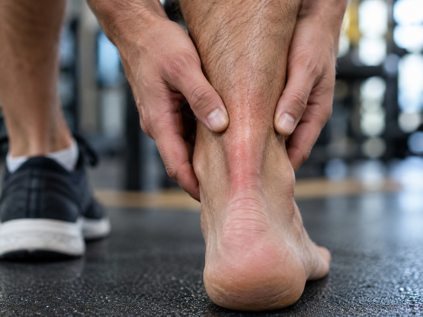
Pathophysiology and Changes in the Achilles Tendon
Surgical analyses of affected tendons show various degenerative changes, including:
Changes in fiber structure and arrangement: Tendon fibers may become disorganized and weakened.
Increase in glycosaminoglycans: These water-binding molecules can retain up to 1,000 times their own weight, which may explain swelling in the tendon.³⁴
The exact cause of tendinopathy remains unclear. Although Achilles tendinopathy is often associated with sporting activities, the condition also frequently occurs in people who do not participate in sports. Overloading of the tendon is the main cause. Mild degeneration may be present without pain, but pain develops when the tendon is overloaded. The condition is usually not caused by trauma.⁵⁶
Terminology
The currently recommended term for this patient group is tendinopathy. This is consistent with updated concepts explaining tendon pathology, including the Tendon Continuum model developed by Cook and Purdum.⁷
The Tendon Continuum Model
The model describes three stages of tendinopathy based on the degree of disorganization and changes in the tendon:
Reactive tendinopathy: An early response to increased load, resulting in reversible cell proliferation and temporary swelling.
Tendon dysrepair: More advanced changes involving increased disorganization of the tendon fibers, although the condition remains partly reversible.
Degenerative tendinopathy: Long-standing changes with irreversible degeneration of tendon structure, including apoptosis (cell death), vascular changes, and fatty infiltration.
Dynamics of the Model
The condition of the tendon can move both up and down within this model depending on the load applied or reduced, particularly in the early stages of tendinopathy. This flexibility in the model emphasizes the importance of individually tailored treatment and load management.
Location of Achilles Tendinopathy
Achilles tendinopathy can be divided into two main types based on location:
Insertional tendinopathy: Occurs at the junction between the Achilles tendon and the calcaneus (less than 2 cm from the insertion).
Midportion tendinopathy: Located within the body of the tendon, more than 2 cm from the tendon insertion.²
Clinically Relevant Anatomy
The Achilles Tendon
The Achilles tendon is the largest and strongest tendon in the human body. It has an enormous capacity to withstand high tensile forces and originates from the distal merging of the gastrocnemius and soleus muscles, inserting distally onto the calcaneus.
Structure and Composition
The structure of the Achilles tendon is key to its strength and function:
Cellular component: Consists of tenocytes and tenoblasts that synthesize all components of the extracellular matrix.
Extracellular matrix: Contains bundles of type I collagen and elastin, with type I collagen being responsible for the tendon's strength.
Ground substance: Located between the collagen fibers and consists of proteoglycans and glycosaminoglycans, which provide structure and elasticity.
Paratenon
The Achilles tendon is surrounded by the paratenon, which functions as an elastic sheath around the tendon. This allows free movement between the tendon and surrounding tissues.
The paratenon consists of loose connective tissue and is found around extra-articular tendons without a synovial sheath, such as the Achilles tendon.
It provides blood supply from nearby blood vessels at several levels.⁹¹⁰
Blood Supply
Blood supply throughout the length of the Achilles tendon is generally poor, which is explained by a low number of blood vessels per cross-sectional area.
The region 4–6 cm above the calcaneus has particularly poor vascularity, which may explain the slow healing rate after trauma.¹¹¹²
The limited blood supply presents a challenge in injuries and contributes to the chronic nature of tendinopathy.
Etiology of Achilles Tendinopathy
Reactive Tendinopathy
The first stage of the tendon pathology continuum is reactive tendinopathy, which is a non-inflammatory proliferative response in the cellular matrix resulting from compressive or tensile overload.
Systematic overloading of the Achilles tendon beyond its physiological limit during physical activity can lead to microtrauma.
Repeated microtrauma, combined with non-uniform tension between the gastrocnemius and soleus, creates frictional forces between the fibers and abnormal concentration of load within the tendon.
This can result in:
Inflammation of the tendon sheath
Degeneration of tendon tissue
Or a combination of both.
Without sufficient recovery time, this can lead to the development of tendinopathy.¹³
Other Risk Factors
Factors that may contribute to chronic overuse injuries and tendon degeneration include:
Reduced arterial blood flow
Local hypoxia
Reduced metabolic activity and nutrition
Persistent inflammatory response
Overpronation
One of the most significant malalignments is overpronation of the foot, which is often associated with Achilles tendinopathy.
Overpronation can increase load on the tendon, potentially increasing the risk of injury.
Acute and Chronic Injuries
Acute tendinopathy:
Often occurs as a result of overuse, blunt trauma, or acute muscular fatigue.
Characterized by an inflammatory response and edema formation.
Incorrect or insufficient treatment may lead to fibrin formation and adhesions.
Chronic tendinopathy:
If the tendon is not unloaded in time, reactive tendinopathy may progress to tendon dysrepair, where increased protein production leads to collagen separation and disorganization in the cellular matrix.
Degenerative tendinopathy is the final stage of the continuum and involves irreversible changes with a poor prognosis.
This condition is often accompanied by peritendinous adhesions, although this does not necessarily mean that one causes the other.
Genetic and Biological Risk Factors
Age, body fat, and waist circumference:
Older men with a higher proportion of android fat mass and a waist circumference above 83 cm have an increased risk of Achilles tendinopathy.¹⁴¹⁵
Genetic predisposition:
Variations in the COL5A1 gene, which regulates production of tendon proteins, have been found to increase the risk of tendinopathy.¹⁶¹⁷
Pain sensitivity:
Patients with chronic Achilles tendinopathy show signs of both peripheral and central pain sensitization.¹⁹²⁰
Recent Research
Age and body composition:
Older men with a high proportion of android fat mass and a waist circumference above 83 cm have an increased risk of Achilles tendinopathy.¹⁴¹⁵
Genetic factors:
Variations in the COL5A1 gene, which is responsible for production of tendon proteins, increase the risk of tendinopathy.¹⁶¹⁷
Pain sensitization:
Chronic Achilles tendinopathy is associated with both peripheral and central pain sensitization.¹⁹²⁰
Implications for Physiotherapy
Treatment of chronic Achilles tendinopathy should include strategies that take pain sensitization into account and address both physical and neurobiological factors to achieve optimal functional recovery.
In other words:
Tendinopathies can develop as a result of multiple factors, including overuse, poor circulation, reduced flexibility, sex, as well as endocrine or metabolic factors. These factors disrupt tendon structure through repeated loading, often eccentric, which creates microtrauma in the tissue.
The process:
Repeated loading: Causes collagen fibers to slide past one another, breaks cross-links, and leads to tissue denaturation.
Inflammation: Tissue damage activates inflammatory processes.
Microtrauma: Accumulating microtrauma weakens collagen cross-links and affects the non-collagenous matrix and vascular elements of the tendon.
These processes disrupt the biomechanical integrity of the tendon, which may ultimately lead to tendinopathy. This chronic condition results from impaired healing capacity and progression of degenerative changes in the tendon.⁶
Risk Factors
There are several known risk factors associated with the development of Achilles tendinopathy. These include:
Obesity: Increased body weight loads the tendon and may increase the risk of injury.
High blood pressure: Associated with reduced circulation and tendon health.
Rapid increase in load: Sudden changes in activity level may exceed the tendon's capacity to adapt.
Type II diabetes: Affects tendon structure and the healing process.
Long-term steroid use: May weaken tendon integrity.
Family history of tendinopathy: Genetic predisposition may play a role.
Other factors:
Poorly fitted footwear.
Age-related changes in tendon tissue.
Clinical Presentation
Morning pain is a key symptom of Achilles tendinopathy. This is because the tendon must tolerate full range of motion and stretch immediately after waking. Symptoms are usually localized to the tendon and surrounding area.
Other symptoms:
Swelling and pain: Less common, but may occur.
Changes in tendon contour: The tendon may become thicker in both the anteroposterior (A-P) and mediolateral (M-L) directions.²¹
In tendinosis:
Tender areas combined with intratendinous swelling that moves with the tendon.
Increased or decreased sensitivity when pressure is applied.
Predictive value for tendinosis in such cases.²²
Observations in the Affected Tendon:
Larger diameter compared with the uninjured side.
Greater stiffness and lower elasticity in the affected tendon.
This clinical picture helps distinguish tendinopathy from other conditions and guides treatment selection.
Differential Diagnoses
To distinguish Achilles tendinopathy from other conditions, the following should be considered:
Plantar fasciitis
Calcaneal stress fracture
Heel pad syndrome: Deep pain in the center of the heel, often resulting from bruising.
Haglund deformity: A prominent calcaneus that may cause bursitis between the calcaneus and the Achilles tendon.
Sever's disease: Irritation of the growth plate at the back of the calcaneus, common in children and adolescents during growth spurts.
Posterior ankle impingement
Medial tendinopathy
Retrocalcaneal bursitis
Sural neuropathy
Lumbar radiculopathy
Ankle osteoarthritis
Deep vein thrombosis
Partial rupture of the Achilles tendon [21].
Examination
Subjective Assessment
A thorough medical history often provides important clues about the mechanism of injury and course of the condition. Questions should identify:
Pain located 2–6 cm proximal to the tendon insertion, with gradual onset.
Pain on palpation, which is characteristic of Achilles tendinopathy [23].
Objective Examination
A comprehensive assessment of the lower extremity is essential. The evaluation includes:
Observation
Assess for muscle atrophy, swelling, asymmetry, joint effusion, and erythema.
Muscle atrophy may indicate the duration of the condition and is often present in chronic cases.
Joint effusion is uncommon in tendinopathy and may indicate intra-articular pathology.
Range of motion, strength, and flexibility
Often reduced on the affected side [24][22].
Palpation
Localized tenderness corresponding to the pain experienced by the patient during activity [25].
Nodules and tendon thickening may be palpable.
Anatomical deformities
For example forefoot deformities, heel varus, excessive pes planus, or foot pronation.
Special Tests
Arc sign and Royal London Hospital test:
These tests may be useful for confirming the diagnosis.
(Refer to the available videos for demonstrations of the tests.)
Functional Tests
Hop tests and heel-rise tests can be used to assess the patient's functional status and document findings over time.
Measurements
Ankle dorsiflexion.
Subtalar range of motion.
Plantar-flexion strength and endurance.
Static arch height, forefoot position, and pain on palpation [23].
Imaging
Although imaging is usually not necessary to diagnose Achilles tendinopathy, it can be useful for differential diagnosis:
Ultrasound: First-line modality for assessing:
Tendon width.
Changes in water content.
Collagen integrity and possible bursitis.
MRI: Used if the diagnosis is uncertain or symptoms are atypical.
Shows increased signal in the Achilles tendon [21].
Advanced Technologies:
Ultrasound elastography and ultra-high-field MRI (UHF MRI):
Useful for assessing the mechanical, structural, and biomechanical properties of the tendon [29].
By combining a thorough clinical evaluation with imaging when indicated, an accurate diagnosis can be established, which is essential for effective treatment.
Outcome Measures
Patient-Reported Outcome Measures
General Lower-Extremity Functional Measures:
Example: Lower Extremity Functional Scale (LEFS). This is not specific to Achilles tendinopathy but provides a comprehensive assessment of function.
Achilles Tendinopathy-Specific Questionnaires:
Example: VISA-A (Victorian Institute of Sport Assessment-Achilles). Measures pain, stiffness, and function specifically related to Achilles tendinopathy [21][30].
Function-Specific Measurement Tools
How much weight the patient can bear on a plantar-flexed foot before pain occurs.
Number of heel raises before onset of pain.
Number of heel drops before onset of pain.
Number of heel drops with a specified weight in a backpack before pain occurs.
How far the patient can walk or run before pain occurs.
Recommended Tools for Clinicians
Use VISA-A to evaluate pain and stiffness.
Use either the Foot and Ankle Ability Measure (FAAM) or Lower Extremity Functional Scale (LEFS) to assess activity and participation in patients with Achilles tendinopathy [23].
Physiotherapy Treatment
Management of Tendinopathy
Patient Education and Advice:
Explain the importance of activity modification.
Avoid complete rest for patients with non-acute Achilles tendinopathy.
Advise patients to continue recreational activity within their pain limits alongside rehabilitation.
Key Elements of Patient Education:
1. Physiotherapy Principles:
Explain the role of mechanical loading in rehabilitation.
2. Modifiable Risk Factors:
For example, body mass index (BMI) and footwear choices.
3. Time Frame for Symptom Improvement:
Inform the patient that improvement may take time, but rehabilitation is a long-term investment in function.
Decision-Support Tools
Achilles Tendinopathy Toolkit:
An evidence-based tool for clinical decision-making, developed to support the management of Achilles tendinopathy.
Effective treatment requires a combination of patient education, individualized goals, and a gradual loading approach that respects the patient's pain threshold.
Optimizing Biomechanics
Comprehensive Biomechanical Assessment
People with Achilles tendinopathy should undergo a comprehensive biomechanical assessment. BC Physical Therapy Tendinopathy Task Force [31] suggests that:
There is limited clinical evidence supporting the use of orthoses in the acute phase.
There is moderate clinical evidence supporting the use of orthoses in the chronic phase.
Clinical recommendations:
In the acute phase: Consider taping as a temporary strategy before assessing the need for orthoses.
In the chronic phase: Orthoses may be considered as an aid to optimize load distribution.
Controlled Tendon Loading
The Importance of Activity
Activities that reduce stress on the tendon should be encouraged, but complete immobilization should be avoided to prevent atrophy [32][5].
Recommendation from the BC Physical Therapy Tendinopathy Task Force [31]:
There is substantial clinical evidence supporting the use of exercise in the chronic phase, although the precise parameters for optimal effectiveness remain unclear.
Eccentric training has particularly strong support, but newer research also shows that heavy slow resistance training (HSR) may be equally effective.
Effects of Strength Training
Strength training related to functional tasks not only reduces tendon pain but also improves excitatory and inhibitory muscle control, which may positively influence tendon loading [33].
Eccentric training has been a popular and effective method over the past decade and is considered the primary nonsurgical treatment for Achilles tendinopathy [34].
Varied Exercise Regimens
There is limited evidence supporting eccentric training alone as the most effective method.
Recent systematic reviews suggest that well-conducted studies comparing different loading programs are lacking [34].
Newer approaches such as isolated concentric training, heavy slow resistance training (HSR) and combined eccentric-concentric training have been proposed but currently lack strong scientific support [35].
Treatment Goals for Tendinopathy
The primary goal is to improve the tendon's energy-storage capacity. This involves increasing the ability of the tendon and associated muscles to handle load and function as a "spring" that stores and releases energy.
Three Key Exercise Types
Isometric Loading: Increases tendon stability and reduces pain.
Isotonic Loading: Builds strength through controlled movements.
Energy-Storage Loading: Mimics functional activities that require energy storage and release in the tendon [36].
For detailed exercise programs and guidance, use of the Achilles Tendinopathy Toolkit: Section D - Exercise Programs is recommended.
Phase 1: Isometric Loading – Achilles Tendon Holds
Changes in the Treatment of Achilles Tendinopathy
Treatment of Achilles tendinopathy has changed considerably in recent years. One of the most important developments is the introduction of isometric loading as a central component of treatment. Isometric loading has been shown to have pain-relieving effects on tendons while maintaining a degree of baseline strength.
Performing Isometric Exercises
Adapted to symptoms and irritability:
For highly irritable (reactive) Achilles tendinopathy, double-leg isometric holds are recommended, often with shorter hold times and fewer repetitions.
For less irritable conditions, single-leg holds may be considered to increase the load.
Position of the isometric hold:
Mid-range: Halfway up onto the toes.
End range: Fully up onto the toes.
Benefits of Isometric Loading
Pain relief: Reduces immediate pain by decreasing pain-related tendon signaling.
Maintains strength: Preserves baseline strength without causing additional irritation.
Flexible adaptation: Can easily be modified based on the patient's symptoms and stage of rehabilitation.
This approach provides an effective starting point for treatment and lays the foundation for progression to more demanding and functional exercises in later phases of rehabilitation [37].
Phase 2: Isotonic Loading – Calf Raises
When Should Isotonic Loading Begin?
Isotonic exercises can begin once both pain inte
nsity and tendon irritability have decreased. There are no absolute rules for when a patient should begin isotonic loading as part of rehabilitation for Achilles tendinopathy. General recommendations include:
Pain reduction: The patient should have less than 5/10 pain on the Numeric Rating Scale (NRS), or tolerable pain during repeated single-leg calf raises.
Reduced stiffness: Morning stiffness in the tendon should be substantially reduced.
Purpose of Isotonic Training
The primary goal is to develop strength in both the Achilles tendon and surrounding musculature, particularly the soleus and gastrocnemius. Adding heavy loads is essential because repeated low loading, such as during walking or running, does not provide sufficient adaptation of the tendon matrix or increase the work capacity of the muscle-tendon unit.
Performing the Exercises
Seated calf raises:
Begin with gradually increasing load.
Perform each repetition over 3–6 seconds to achieve sufficient tension development in the tendon.
Standing calf raises:
Performed through the middle portion of the muscle's range of motion to avoid compressive loading of the tendon.
Heavy slow resistance (HSR) repetitions through the mid-range minimize irritation that may occur with compression:
At end-range plantar flexion (fully raised onto the toes).
During dorsiflexion (heel lowered below the level of a step).
Benefits of Isotonic Loading
Strengthens the tendon: Increases the tendon matrix's capacity to tolerate load.
Improves muscle strength: Particularly in the soleus and gastrocnemius.
Reduces pain: Promotes adaptation that may reduce pain over time.
Prepares for functional movements: Increases the work capacity of the muscle-tendon unit.
This phase is a critical step in rehabilitation, and progression should be tailored according to the patient's pain, tolerance, and functional level [38][39].
Phase 3: Energy Storage – Plyometric Exercises
Importance of Plyometric Exercises
The final and crucial phase of rehabilitation for Achilles tendinopathy involves energy-storage exercises. These exercises focus on restoring the tendon's ability to absorb and release energy through the stretch-shortening cycle. This is the mechanism activated when a person lands after a jump and then pushes off through the toes.
When Can These Exercises Begin?
Plyometric exercises can be initiated when the following criteria are met:
Minimal morning stiffness: The patient reports minimal or noticeably reduced Achilles tendon stiffness on waking.
Progress with isotonic exercises: The patient has demonstrated good progression with calf-raise exercises and heavy slow resistance training.
Minimal tenderness: Only mild tenderness on palpation of the tendon.
Running tolerance: The patient can perform light jogging without a flare-up of symptoms or increased tendon irritability.
Purpose of Plyometric Exercises
Plyometric exercises:
Improve the elastic properties of the tendon.
Increase force production through rapid transition between eccentric and concentric muscle action.
Prepare the tendon and muscles for sporting activities that require explosiveness and rapid movements.
Examples of Exercises
Double-leg jumps:
Perform repeated jumps with an emphasis on even push-off and controlled landing.
Single-leg jumps:
Begin with short distances and progress to longer jumps as strength improves.
Single-leg bounding:
Perform explosive bounds, emphasizing powerful push-off.
Band-resisted jumping:
Use an elastic resistance band to add resistance during the jumping movement. This challenges stability and the tendon's ability to tolerate load.
Progression and Adjustment
Increase in load: Time under tension can gradually be increased during heavy resistance training to promote tendon adaptation.
Speed training: Increasing the speed of plyometric exercises may improve explosiveness and prepare the tendon for sporting activities requiring high loads in the stretch-shortening cycle.
This phase is essential for ensuring that the patient can safely return to high-intensity activities and sport [40].
Adjunctive Treatments
In addition to approaches that optimize biomechanics and include exercise programs, adjunctive treatments may be used as support. These therapies are mainly intended for symptom relief, rather than to resolve or prevent the injury itself.
Manual Therapy
Manual therapy can be used in both acute and chronic stages of Achilles tendinopathy, but with different purposes and limitations:
Acute stage:
There is no extensive clinical evidence, but expert consensus suggests that joint mobilization/manipulation may be useful if the examination identifies joint restrictions.
For example, ankle mobilization may improve dorsiflexion at the talocrural joint or correct varus and valgus restrictions at the subtalar joint.
Chronic stage:
There is some clinical evidence and stronger expert consensus supporting the use of joint mobilization/manipulation in chronic tendinopathy when joint restrictions are identified.
Assessment of the hip, knee, foot, and ankle should be included to identify joint dysfunction that may contribute to pain.
Transverse Friction Massage
The effect of deep transverse friction massage has not been scientifically established and generally produces limited results.
In the chronic stage, there is some clinical evidence supporting the use of soft-tissue techniques such as friction massage.
Friction massage may be considered as part of treatment after careful evaluation, but should be viewed as an adjunct to a comprehensive treatment plan.
Important Considerations
Manual therapy and soft-tissue techniques are most effective when used following a thorough examination that identifies specific biomechanical restrictions.
These therapies are not a substitute for structured rehabilitation, but they may help reduce pain and improve range of motion in some patients.
These approaches may be combined with other treatment strategies to improve the patient's overall function and quality of life [31][41][42][43].
Electrotherapy and Related Modalities
Extracorporeal Shockwave Therapy (ESWT)
There is conflicting evidence for the use of ESWT in the chronic phase of Achilles tendinopathy. Outcomes appear to depend more on the dose of shockwave energy (EFD - energy flux density = mJ/mm²) than on the type of shockwave generation (focused vs radial ESWT). Use of anesthesia in high-energy protocols may reduce the effectiveness of ESWT.
Recommendations for ESWT:
Low-energy protocols without anesthesia are recommended because they are more practical, tolerable, and cost-effective while providing comparable outcomes.
Parameters for low-energy ESWT:
EFD: 0.18–0.3 mJ/mm² (2–4 Bars)
Number of shocks: 2000–3000
Frequency: 15–30 Hz
Number of sessions: 3–5, at weekly intervals
Ultrasound and Low-Level Laser Therapy (LLLT)
There is no clinical evidence supporting the use of ultrasound or LLLT in the treatment of Achilles tendinopathy.
Iontophoresis
A small amount of evidence supports the use of iontophoresis with dexamethasone in the acute phase, but not in the chronic phase. This modality is still being investigated.
Recommendations for iontophoresis in the acute phase:
Dexamethasone (water-based): 0.4%
Dose: 80 mA-min
Number of sessions: 6 sessions over 3 weeks
Combined with concentric-eccentric exercise if the exercise load is tolerated.
Taping
Anti-pronation taping: Supported by expert opinion but not by clinical evidence. It may be considered as a temporary solution, particularly in the acute phase.
Rigid taping: May be used to reduce load on the Achilles tendon and/or alter foot posture.
Elastic taping: Should not be used to reduce pain or improve functional performance in Achilles tendinopathy.
Night Splints
Acute phase: Expert opinion supports the use of night splints and orthoses.
Chronic phase: There is moderate evidence against the use of night splints in the chronic phase.
They may be considered in the acute phase, but should be avoided in the chronic phase and combined with exercise.
Dry Needling
Dry needling combined with injection (ultrasound-guided) and eccentric training may be considered for patients with symptoms lasting more than three months and increased tendon thickness. This approach may help reduce pain and improve functional capacity.
Medical Treatment
Pharmacological Treatment
Inflammation is a necessary part of the body's repair process in injured tissue. Use of certain medications, such as corticosteroids and quinolones, may inhibit inflammation and thereby delay or disrupt the repair process. Even in patients who do not use these medications, tendinopathy may arise from a disturbed repair process. This highlights the importance of balanced and appropriate management of these conditions. [44]
Corticosteroid Injections (CSI)
Corticosteroid injections may provide short-term pain relief but have shown no benefit or negative effects in the longer term.
Benefits:
Short-term improvement in pain during walking.
Reduction in tendon thickness measured by ultrasound.
Limitations and Contraindications:
Intratendinous injection is contraindicated because of catabolic effects that may weaken tendon structure.
Peritendinous injections have fewer adverse effects and may be considered as an adjunct to a carefully planned treatment program.
Clinical use:
Corticosteroid injections may be most useful for relieving pain during a period in which the patient is following a structured exercise program. Long-term improvements are limited, however. [45]
Sclerotherapy
Sclerotherapy focuses on treating neovascularization, which may contribute to tendon pain. A vascular sclerosant such as Polidocanol (a nitrogen-free substance with sclerosing and anesthetic effects) is injected into areas of neovascularization anterior to the tendon.
Study results:
At short-term follow-up (6 months), the majority of patients reported being pain-free after an average of two treatments.
At 2-year follow-up, patients remained pain-free, and neovascularization was not detected on ultrasound.
A reduction in tendon thickness and an almost normal tendon structure were observed.
Rehabilitation after sclerotherapy:
1–3 days: Complete rest.
After 2 weeks: Gradual increase in load, including jumping, fast running, and heavy strength training.
Maximum loading should be avoided during the first weeks.
Clinical role:
Sclerotherapy may be an option for patients who do not respond to eccentric training. This suggests a potential role in the treatment of chronic tendinopathy. [45]
Platelet-Rich Plasma (PRP) Injections
Research shows that injections of Platelet-Rich Plasma (PRP) over a period of three months in people with chronic Achilles tendinopathy do not have a significant positive effect compared with placebo (saline). The only observed difference between PRP and placebo was an increase in tendon thickness, suggesting that PRP may contribute to increased tendon thickness compared with saline injections. This increase in thickness may be of interest but has limited clinical relevance without other positive effects on symptoms or function. [45]
Minimally Invasive Procedures
Minimally invasive procedures have shown effectiveness in the treatment of insertional Achilles tendinopathy, a common chronic musculoskeletal condition characterized by pain at the Achilles tendon. A recent study highlights percutaneous Zadek osteotomy (ZO) as a keyhole technique that may provide significant pain relief.
Benefits of ZO:
Reduces pain within six weeks, compared with 23 weeks for traditional open surgery.
Shorter rehabilitation period:
0–2 weeks: Protect the foot with a splint or walker boot.
2–6 weeks: Gradual weight-bearing with a removable walker boot.
Physiotherapy begins two weeks after surgery.
Patients may wear regular shoes six weeks after the procedure.
The procedure is particularly useful for athletes who perform powerful push-off movements, such as basketball and football players. [46]
Operative Surgery
The primary goal of surgical treatment for Achilles tendinopathy is to stimulate a chemically mediated response in the tendon by irritating the tissue. This can be achieved through different surgical approaches, including:
Simple procedures: Minimal intervention to create a local response.
Percutaneous tenotomy: A needle-based technique used to release or irritate the tendon.
Open surgery: Used for more severe cases and involves removal of damaged tendon tissue.
Effectiveness:
75% of patients who underwent tenotomy reported positive outcomes after 18 months.
Open surgery has shown better outcomes in cases where the tendon does not have focal lesions, suggesting that the technique may be more appropriate for diffusely degenerative tendon tissue.
Although surgical procedures can provide good outcomes, they should be considered a last resort after unsuccessful conservative treatment. [45]
Conclusion
The choice of treatment, whether PRP, minimally invasive procedures, or surgery, depends on the patient's symptoms, activity level, and response to conservative measures. Physiotherapy rehabilitation is an integral part of any treatment plan to ensure optimal function and reduce the risk of recurrence.
Sources:
1. Clain MR, Baxter DE. Achilles tendinitis. Foot & Ankle. 1992 Oct;13(8):482-7.
2. Radiopaedia. Achilles Tendinopathy. Available from: https://radiopaedia.org/articles/achilles-tendinopathy (accessed 10.6.2022).
3. Schubert TE, Weidler C, Lerch K, Hofstädter F, Straub RH. Achilles tendinosis is associated with sprouting of substance P positive nerve fibres. Annals of the Rheumatic Diseases. 2005 Jul 1;64(7):1083-6.
4. Verywell Health. The Health Benefits of Glycosaminoglycans. Available from: https://www.verywellhealth.com/glycosaminoglycans-5092414 (accessed 11.6.2022).
5. Wilson JJ, Best TM. Common overuse tendon problems: a review and recommendations for treatment. American Family Physician. 2005 Sep 1;72(5):811-8.
6. Paavola M, Kannus P, Järvinen TA, Khan K, Józsa L, Järvinen M. Achilles tendinopathy. JBJS. 2002 Nov 1;84(11):2062-76.
7. Cook JL, Purdam CR. Is tendon pathology a continuum? A pathology model to explain the clinical presentation of load-induced tendinopathy. British Journal of Sports Medicine. 2009 Jun 1;43(6):409-16.
8. Van der A, Van de Walle L. Het excentrisch trainen van de kuitspieren.
9. Radiopaedia. Paratenon. Available from: https://radiopaedia.org/articles/paratenon (accessed 10.6.2022).
10. Kiewiet NJ, Holthusen SM, Bohay DR, Anderson JG. Gastrocnemius recession for chronic noninsertional Achilles tendinopathy. Foot & Ankle International. 2013 Apr;34(4):481-5.
11. Ahmed IM, Lagopoulos M, McConnell P, Soames RW, Sefton GK. Blood supply of the Achilles tendon. Journal of Orthopaedic Research. 1998 Sep;16(5):591-6.
12. Carr AJ, Norris SH. The blood supply of the calcaneal tendon. The Journal of Bone and Joint Surgery. British Volume. 1989 Jan;71(1):100-1.
13. Young JS, Maffulli N. Etiology and epidemiology of Achilles tendon problems. The Achilles Tendon. 2007 Sep 15:39-49.
14. Gaida JE, Alfredson H, Kiss ZS, Bass SL, Cook JL. Asymptomatic Achilles tendon pathology is associated with a central fat distribution in men and a peripheral fat distribution in women: a cross-sectional study of 298 individuals. BMC Musculoskeletal Disorders. 2010 Dec;11(1):1-9.
15. Gaida JE, Ashe MC, Bass SL, Cook JL. Is adiposity an under‐recognized risk factor for tendinopathy? Arthritis Care & Research. 2009 Jun 15;61(6):840-9.
16. Mokone GG, Schwellnus MP, Noakes TD, Collins M. The COL5A1 gene and Achilles tendon pathology. Scandinavian Journal of Medicine & Science in Sports. 2006 Feb;16(1):19-26.
17. September AV, Cook J, Handley CJ, van der Merwe L, Schwellnus MP, Collins M. Variants within the COL5A1 gene are associated with Achilles tendinopathy in two populations. British Journal of Sports Medicine. 2009 May 1;43(5):357-65.
18. Abate M, Schiavone C, Salini V, Andia I. Occurrence of tendon pathologies in metabolic disorders. Rheumatology. 2013 Apr 1;52(4):599-608.
19. Ames PR, Longo UG, Denaro V, Maffulli N. Achilles tendon problems: not just an orthopaedic issue. Disability and Rehabilitation. 2008 Jan 1;30(20-22):1646-50.
20. Eckenrode BJ, Kietrys DM, Stackhouse SK. Pain sensitivity in chronic Achilles Tendinopathy. International Journal of Sports Physical Therapy. 2019 Dec;14(6):945.
21. Cook JL, Khan KM, Purdam C. Achilles tendinopathy. Manual Therapy. 2002 Aug 1;7(3):121-30.
22. Luscombe KL. Achilles tendinopathy. Trauma. 2003;215-225.
23. Martin RL, Chimenti R, Cuddeford T, Houck J, Matheson JW, McDonough CM, et al. Achilles pain, stiffness, and muscle power deficits: Midportion Achilles tendinopathy revision 2018. Journal of Orthopaedic & Sports Physical Therapy. 2018 May;48(5):A1-38.
24. Hammer WI, editor. Functional Soft-Tissue Examination and Treatment by Manual Methods. Jones & Bartlett Learning; 2007.
25. Shibuya N, Thorud JC, Agarwal MR, Jupiter DC. Is calcaneal inclination higher in patients with insertional Achilles tendinosis? The Journal of Foot and Ankle Surgery. 2012 Nov 1;51(6):757-61.
26. Royal London Hospital Test (CR). Available from: https://www.youtube.com/watch?v=nM3yu_TR4H8&t=2s.
27. CRTechnologies. Arc Sign Achilles. Available from: https://www.youtube.com/watch?v=zhP0CAsQr7U&t=24s.
28. Fouré A. New imaging methods for non-invasive assessment of mechanical, structural, and biochemical properties of human Achilles tendon: A mini review. Frontiers in Physiology. 2016 Jul 27;7:324.
29. Robinson JM, Cook JL, Purdam C, et al. The VISA-A questionnaire: A valid and reliable index of the clinical severity of Achilles tendinopathy. British Journal of Sports Medicine. 2001 Oct 1;35(5):335-41.
30. BC Physical Therapy Tendinopathy Task Force. Achilles Tendinopathy Toolkit. UBC Department of Physical Therapy. 2012.
31. Scott A, Huisman E, Khan K. Conservative treatment of chronic Achilles tendinopathy. CMAJ. 2011 Jul 12;183(10):1159-65.
32. Rio E, Kidgell D, Moseley GL, et al. Tendon neuroplastic training: Changing the way we think about tendon rehabilitation. British Journal of Sports Medicine. 2016 Feb 1;50(4):209-15.
33. Malliaras P, Barton CJ, Reeves ND, Langberg H. Achilles and patellar tendinopathy loading programmes. Sports Medicine. 2013 Apr 1;43(4):267-86.
34. Beyer R, Kongsgaard M, Hougs Kjær B, et al. Heavy slow resistance versus eccentric training as treatment for Achilles tendinopathy: A randomized controlled trial. The American Journal of Sports Medicine. 2015 Jul;43(7):1704-11.
35. Cook JL, Purdam CR. The challenge of managing tendinopathy in competing athletes. British Journal of Sports Medicine. 2014 Apr 1;48(7):506-9.
36. Single leg heel raise isometric hold. Available from: https://www.youtube.com/watch?v=7Udb4E3Uks8.
37. Soleus calf raises-seated. Available from: https://www.youtube.com/watch?v=C_UwdAuD7ZY.
38. Single leg heel raise isometric hold. Available from: https://www.youtube.com/watch?v=qW2XAz8hYf0&feature=emb_logo.
39. Achilles Tendonitis Rehab Phase 3. Available from: https://www.youtube.com/watch?v=t9IwvBjQA8I.
40. Stasinopoulos D, Stasinopoulos I. Comparison of effects of exercise programme, pulsed ultrasound, and transverse friction in the treatment of chronic patellar tendinopathy. Clinical Rehabilitation. 2004 Jun;18(4):347-52.
41. Joseph MF, Taft K, Moskwa M, Denegar CR. Deep friction massage to treat tendinopathy: A systematic review. Journal of Sport Rehabilitation. 2012 Nov 1;21(4):343-53.
42. Alfredson H, Cook J. A treatment algorithm for managing Achilles tendinopathy: New treatment options. British Journal of Sports Medicine. 2007;41(4):211-216.
43. Silbernagel KG, Hanlon S, Sprague A. Current clinical concepts: Conservative management of Achilles tendinopathy. Journal of Athletic Training. 2020 May;55(5):438-47.


