

Scapholunate Dissociation
- Fysiobasen

- Dec 21, 2025
- 6 min read
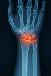
Scapholunate dissociation (Scapholunate Dissociation, SLD) is the most common and clinically important ligament injury in the wrist. It occurs due to injury to the scapholunate ligament (SLL) and leads to instability between the scaphoid and lunate. Radiologically, this appears as an abnormally large gap between the bones – often referred to as the "Terry Thomas sign"¹–³.
Anatomy and Biomechanics
The scapholunate ligament is C-shaped and consists of three parts: volar, membranous, and dorsal. The dorsal portion is the strongest (260 N) and the most important stabilizer. The volar portion contributes to rotational stability (118 N), while the membranous middle portion is weak and avascular⁴.
Primary load paths through the wrist:
50% of axial load passes via the radioscaphoid joint
35% via the radiolunate joint
The injury often occurs because the scaphoid and lunate are forced in different directions under load. Secondary stabilizers include the scaphotrapeziotrapezoid ligament, scaphocapitate ligament, and dorsal intercarpal ligament².
Dynamic stabilizers include:
APL, ECRL, and FCU (via midcarpal supination)
FCR contributes to scaphoid flexion and capitate pronation
Epidemiology and Etiology
Scapholunate instability is common after trauma – especially after a fall on an outstretched hand (FOOSH) with the wrist in extension and ulnar deviation. This produces an asymmetric force transmission between scaphoid and lunate⁵.
5% of all wrist sprains involve concomitant SL injury
13.4% of distal radius fractures are associated with SL injury – particularly chauffeur fractures and nondisplaced scaphoid fractures³
Overuse injuries also occur, for example with crutch use, prolonged static loading, or repetitive wrist extension².
Clinical Presentation
Patients often report:
Fall or trauma to the wrist in extension and ulnar deviation
Pain in the dorso-radial wrist, sometimes in the snuffbox region or scaphoid tubercle
Clicking, giving way, reduced grip strength, and swelling
Pain with loading, rotation, and gripping
Often, the patient initially has normal radiographs, and symptoms may develop over months.
Radiological Classification and Stages
Watson et al. defined four stages of instability based on radiographs⁸:
Pre-dynamic: Symptoms without visible damage
Dynamic instability: Pathological widening visible only on stress images
Static SL dissociation: Visible SL gap > 3 mm and SL angle > 70°
SLAC (Scapholunate Advanced Collapse): Progressive osteoarthritis
Radiological features:
PA radiograph: scapholunate gap > 3 mm (= pathological)
Lateral radiograph: scapholunate angle > 70°
Stress views (clenched fist, ulnar deviation) accentuate findings
MRI and CT are used in unclear cases
SLAC progression:
Osteoarthritis in the radial styloid
Radioscaphoid osteoarthritis
Capitolunate osteoarthritis
Pan-carpal osteoarthritis
Complications
Untreated scapholunate dissociation may lead to two main complications:
Scapholunate Advanced Collapse (SLAC)
Wrist osteoarthritis
SLAC is a progressive, specific form of degenerative arthropathy that develops in several stages, first involving the radial styloid and later radioscaphoid and capitolunate joints. The result is often considerable functional loss of the wrist if untreated³.
Examination
Palpation
Pain in the snuffbox or over the palmar scaphoid tubercle
Tenderness distal to Lister’s tubercle
Special tests
Watson’s test (Scaphoid shift maneuver):
Performed with the wrist in slight extension
Examiner places thumb over the scaphoid tubercle and moves the wrist from ulnar to radial deviation
Positive test: click and pain – sign of dorsal subluxation
Scapholunate Ballottement Test:
Stabilize the lunate and mobilize the scaphoid
Positive test: pain and excessive movement
Imaging
Standard radiographs: AP, lateral, clenched fist
Ulnar deviation to reveal dynamic instability
Important radiological findings:
SL gap > 3 mm (Terry Thomas sign)
SL angle > 60° (indicates dorsal intercalated segment instability – DISI)
Scaphoid ring sign
Disruption of Gilula’s lines
Differential Diagnoses
For ulnar-sided, dorsal, or volar wrist pain, other conditions must be excluded:
Dorsal wrist
SL injury, LTIL injury, DRUJ instability, occult ganglion, Kienböck’s, carpometacarpal boss, PIN irritation
Ulnar wrist
TFCC injury, ulnar impaction, FCU/ECU tendinitis, tendon subluxation
Radial wrist
De Quervain’s, intersection syndrome, scaphoid fracture, cheiralgia paresthetica
Volar wrist
Ganglion, carpal instability, pisotriquetral arthritis, Preiser’s disease
Palmar region
Carpal tunnel, ulnar neuritis, hamate hook fracture, Guyon’s canal compression
Outcome Measures
To assess function and progression in scapholunate dissociation, recommended measures include:
DASH (Disabilities of the Arm, Shoulder and Hand)
PRWE (Patient-Rated Wrist Evaluation)
Medical Treatment
Treatment of scapholunate dissociation (SLD) offers several surgical options, but no clear gold standard exists. Studies and meta-analyses show large variation in technique and outcome reporting, and the surgeon’s experience often outweighs the specific procedure⁷ ⁹ ¹⁶.
Acute injuries (within 4–6 weeks):
Closed reduction + cast (8 weeks)
Closed or open reduction with percutaneous K-wires
Open reduction + direct SLL repair
Open reconstruction with tendon graft
Chronic injuries without arthritis:
Blatt capsulodesis
Scaphoid tenodesis
Tendon reconstruction of SLL
Scaphoid-trapezium-trapezoid fusion
Chronic SLD with arthritis (SLAC):
Proximal row carpectomy
Scaphoidectomy + four-corner fusion
Total wrist arthrodesis
Soft tissue reconstructions provide better biomechanical restoration but less viscoelasticity than the original ligament. Skeletal procedures (e.g. fusions) are more predictable but irreversible. Garcia-Elias et al. developed a treatment algorithm based on stage and patient function¹⁶.
Surgery usually provides good symptom relief, although full function is not always regained. Patients often report improved pain and function compared to conservative management⁹ ¹⁷.
Rehabilitation and Physiotherapy
Acute phase (non-operative):
Managed like an acute ankle ligament injury
Rest, avoid load, supportive bandage or orthosis
NSAIDs, hot/cold packs, contrast baths as needed
Avoid activities stressing the wrist in extension
Chronic phase (non-operative):
Focus on functional limitations: pain, reduced grip strength, reduced range of motion
Orthosis may be used during heavy activity
Patient education and activity modification
Post-surgery:
Cast up to 10 weeks, then gradual mobilization
Orthosis during physical load up to 1 year post-op
Rehabilitation timeline:
Return to activity: 4–6 months
Full function: up to 12–15 months⁴ ²¹
Effective Exercises
Stabilization and neuromuscular control are crucial. A combination of proprioception and strength training provides the best results.
Reactivation of flexor carpi radialis (FCR) for scaphoid stability
Eccentric and concentric strength training with theraband
Hammer/club exercises for pronation and supination
Isometric strength for flexion/extension
“Prayer stretch” and mobilization in prayer position
Grip training with dynamometer – dosed to the contralateral side
Clinical Summary
Scapholunate dissociation is a common but often overlooked complication after FOOSH injuries. Many patients develop secondary conditions such as SLAC and osteoarthritis over time. The most evidence-based treatment is surgical, but no universal solution exists – choice of method should be based on stage and surgeon expertise.
Physiotherapy should be individualized and function-oriented, focusing on mobility, proprioception, and graded loading. Multidisciplinary follow-up provides the best outcomes.
Soruces:
Duke Orthopaedics: Wheeless' Textbook of Orthopaedics. http://www.wheelessonline.com/ortho/scapholunate_instability (accessed 15 October 2011).
Konopka G, Chim H. Optimal management of scapholunate ligament injuries. Orthopedic research and reviews. 2018;10:41.
Andersson JK. Treatment of scapholunate ligament injury: current concepts. EFORT open reviews. 2017 Sep;2(9):382-93.
Van Overstraeten L, Camus EJ, Wahegaonkar A, Messina J, Tandara AA, Binder AC, Mathoulin CL. Anatomical description of the dorsal capsulo-scapholunate septum (DCSS)—arthroscopic staging of scapholunate instability after DCSS sectioning. Journal of wrist surgery. 2013 May;2(02):149-54.
Goelz L, Kim S, Güthoff C, Eichenauer F, Eisenschenk A, Mutze S, Asmus A. ACTION trial: a prospective study on diagnostic Accuracy of 4D CT for diagnosing Instable ScaphOlunate DissociatioN. BMC Musculoskelet Disord. 2021 Jan 15;22(1):84.
Tomas A. Scapholunate Dissociation. Journal of Orthopaedic & Sports Physical Therapy. 2018 Mar;48(3):225-.
Lau S, Swarna SS, Tamvakopoulos GS. Scapholunate dissociation: an overview of the clinical entity and current treatment options. European Journal of Orthopaedic Surgery & Traumatology. 2009 Aug 1;19(6):377-85.
Konopka G, Chim H. Optimal management of scapholunate ligament injuries. Orthopedic research and reviews. 2018;10:41.
Bloom HT, Freeland AE, Bowen V, Mrkonjic L. The Treatment of Chronic Scapholunate Dissociation: An Evidence-Based Assessment of the Literature. Orthopedics. 2003;26(2):195-203
Andersson JK. Treatment of scapholunate ligament injury: current concepts. EFORT open reviews. 2017 Sep;2(9):382-93.
Duke Orthopaedics: Wheeless' Textbook of Orthopaedics. http://www.wheelessonline.com/ortho/scapholunate_advanced_collapse_slac.
Opreanu RC, Baulch M, Katranji A. Reduction and maintenance of scapholunate dissociation using the TwinFix screw. Eplasty. 2009;9.
Watson's Test. http://en.wikipedia.org/wiki/Watson%27s_test. (accessed 24 October 2011).
Imada AO, Welch K, Mlady G, Moneim MSA. The tangential view described by Moneim to demonstrate scapholunate dissociation: an update. Eur J Orthop Surg Traumatol. 2022.
Jacobson MD, Plancher KD. Evaluation of hand and wrist injuriesin athletes. Operative Techniques in Sports Medicine. 1996 Oct 1;4(4):210-26.
Garcia-Elias M, Lluch AL, Stanley JK. Three-ligament tenodesis for the treatment of scapholunate dissociation: indications and surgical technique. The Journal of hand surgery. 2006 Jan 1;31(1):125-34.
Caloia M, Caloia H, Pereira E. Arthroscopic scapholunate joint reduction. Is an effective treatment for irreparable scapholunate ligament tears?. Clinical Orthopaedics and Related Research®. 2012 Apr 1;470(4):972-8.
Wolff AL, Wolfe SW. Rehabilitation for scapholunate injury: application of scientific and clinical evidence to practice. Journal of Hand Therapy. 2016 Apr 1;29(2):146-53.
Capele A, et al. Mayo Clinic Health Letter - Tools for Healthier Lives. 2011;29(1):1-3. Mayo Foundation for Medical Education and Research, 200 first St. SW, Rochester, MN 55905.http://www.businesswire.com/news/home/20110117005129/en/Mayo-Clinic-Health-Letter-January-2011-Reducing Accessed: November 27th, 2011.
Salvà-Coll G, Garcia-Elias M, Llusá-Pérez M, Rodríguez-Baeza A. The role of the flexor carpi radialis muscle in scapholunate instability. The Journal of hand surgery. 2011 Jan 1;36(1):31-6.
Goldberg SH, Strauch RE, Rosenwasser MP. Scapholunate and lunotriquetral instability in the athlete: Diagnosis and management. Operative Techniques in Sports Medicine. 2006 Apr 1;14(2):108-21


