Endocarditis
- Fysiobasen

- Dec 15, 2025
- 5 min read
Endocarditis is an inflammatory condition of the endocardium, the innermost lining of the heart and heart valves. The condition is most often caused by an infection, and the most common form is known as infective endocarditis (IE). Endocarditis can lead to severe complications such as valve destruction, heart failure, and the spread of infection to other organs. Early diagnosis and treatment are crucial to prevent permanent damage¹.

Clinically relevant anatomy
Endocarditis can affect several cardiac structures, particularly the endocardium (the heart’s inner lining) and the heart valves, and in some cases also the myocardium. The valves are especially vulnerable because they lack a direct blood supply and therefore have a reduced ability to combat infection.
Causes and pathophysiology
The most common cause of endocarditis is bacterial infection, most frequently with Staphylococcus aureus and Streptococcus viridans. Fungal and viral infections may also lead to endocarditis, but these are far less common. Infection usually begins when bacteria enter the bloodstream (bacteremia), for example following dental procedures, surgery, or intravenous drug use. The bacteria most easily adhere to damaged endocardium or prosthetic heart valves.
In the infected area, so-called vegetations are formed — clumps of fibrin (a blood-clotting protein), platelets, and microbes. These vegetations can destroy the valves and may detach, allowing small fragments (emboli) to travel through the bloodstream to other organs and cause complications².
In recent decades, the profile of endocarditis has shifted as more patients live longer with valve prostheses or implanted cardiac devices. This has increased the incidence of prosthetic valve endocarditis, which tends to be more complex and more difficult to treat than infections affecting native valves³.
Risk factors
The following factors increase the risk of developing infective endocarditis:
Congenital heart defects
Prosthetic heart valves or prior valve surgery
Previous endocarditis
Cardiac or valve operations
Implanted cardiac devices (e.g., pacemaker, defibrillator)
Intravenous drug use
Dental procedures with bleeding risk
Surgical interventions in airways, urinary tract, gastrointestinal tract, or during skin infections
Hypertrophic cardiomyopathy (thickened heart muscle)
High blood pressure (hypertension)
Age over 50 years (the condition is more common in older adults)
Male sex (men are affected about twice as often as women)⁴
Symptoms and clinical presentation
The most common symptoms of endocarditis include:
Fever, chills, and night sweats (often persisting for several days)
Fatigue, reduced general condition, tiredness
Shortness of breath during activity
Swelling in the legs, feet, or abdomen (signs of heart failure)
Muscle pain and joint pain
New heart murmur (detected by auscultation)
Petechiae (small pinpoint hemorrhages in the skin, e.g., under fingernails – splinter hemorrhages)
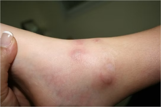
Red, painless spots on the palms and soles (Janeway lesions)
Painful nodules on fingers and toes (Osler’s nodes)
Symptoms may be diffuse and develop gradually, especially in older patients. Severe cases may cause valvular failure, stroke, or abscesses in other organs⁵.
Diagnostics
When endocarditis is suspected, a thorough clinical examination is performed, including auscultation of the heart for new or altered murmurs. Blood tests are taken to detect infection: blood cultures to identify microbes, as well as CRP, ESR, and complete blood count.
An echocardiogram is essential to visualize vegetations, valve damage, and any leakage. Blood tests also guide antibiotic selection and can detect signs of organ damage⁴,⁵.
Treatment and medical management
Endocarditis is a serious condition requiring immediate hospital admission. Treatment includes:
Intravenous antibiotics for 4–6 weeks (choice of drug is based on blood culture)
Surgical treatment is considered in cases of:
Detached vegetation or risk of stroke/embolism
Valvular dysfunction with heart failure
Severe organ damage or uncontrolled infection
In the presence of complications such as myocardial infarction, stroke, or abscesses, treatment is coordinated with specialists⁵.
The prognosis is significantly better with early treatment. If left untreated, endocarditis can lead to brain abscesses, heart failure, severe embolisms, or death.
Prevention
Effective prevention aims to reduce the risk of bacteria entering the bloodstream:
Good oral hygiene and regular dental check-ups are crucial for those in risk groups
Prompt treatment of infections in skin, wounds, or teeth
Good hand hygiene reduces risk
Antibiotic prophylaxis is recommended only for selected high-risk patients undergoing certain procedures, and should not be used routinely to avoid resistance⁴
Prognosis and course
Early diagnosis and treatment provide a good prognosis, but the condition still carries significant mortality and risk of severe complications, especially in older patients or with delayed treatment. Follow-up and monitoring after endocarditis are important to prevent recurrence or to detect long-term complications at an early stage⁵.
Physical Therapy

Physiotherapy plays an important role in rehabilitation after endocarditis, particularly for patients who have experienced complications such as heart failure, reduced physical capacity, or functional decline after prolonged hospitalization. The main tasks of the physiotherapist are to assess physical function, ensure safe mobilization, and support gradual rebuilding of strength, endurance, and activity level¹.
Assessment and evaluation
At the start of rehabilitation, a thorough functional assessment is carried out. This includes evaluation of endurance, muscle strength, balance, mobility, and possible signs of heart failure, such as shortness of breath, edema, or fatigue during activity. Monitoring of respiration, heart rate, and blood pressure during activity is crucial to ensure safe progression.
Interventions and follow-up
The exercise program is tailored individually, based on the patient’s health status, comorbidities, and complications. A gradual increase in activity level is recommended, focusing on:
Early mobilization to prevent muscle weakness and complications after bed rest
Light to moderate endurance training, with monitoring of symptoms
Strength training with cautious progression, particularly in patients with heart failure or low energy
Functional exercises targeting daily activities
Patient education
Education and motivation for an active lifestyle are central. The physiotherapist provides guidance on gradually increasing physical activity, the importance of avoiding inactivity, and how to adapt activity to symptoms and limitations. Many patients need support to overcome fear of activity after serious illness.
Interdisciplinary collaboration
The physiotherapist works closely with physicians and other healthcare professionals to adjust exercise according to medical status. If there are signs of relapse, heart failure, or new infection, the exercise program should be modified, and further medical assessment considered.
Long-term goal
The goal is to help the patient return to their previous level of function, or to achieve the best possible independence and quality of life. For many, participation in a cardiac rehabilitation program will be appropriate, either individually or in group settings.
References
Baddour LM, Wilson WR, Bayer AS, Fowler VG, Tleyjeh IM, Rybak MJ, et al. Infective Endocarditis in Adults: Diagnosis, Antimicrobial Therapy, and Management of Complications: A Scientific Statement for Healthcare Professionals From the American Heart Association. Circulation. 2015;132(15):1435–86.
Murdoch DR. Clinical Presentation, Etiology, and Outcome of Infective Endocarditis in the 21st Century. Arch Intern Med. 2009;169(5):463.
Rajani R, Klein JL. Infective endocarditis: A contemporary update. Clin Med. 2020;20(1):31–5.
NHS. Endocarditis. Available from: https://www.nhs.uk/conditions/endocarditis/ [accessed 05.07.2025]
MedlinePlus. Endocarditis. Available from: https://medlineplus.gov/ency/article/001098.htm [accessed 05.07.2025]


