Sarcoidosis
- Fysiobasen

- Dec 24, 2025
- 4 min read
Sarcoidosis is a potentially life-threatening, multisystem disease of unknown cause with no known cure¹. It is characterized by granulomatous inflammation that can affect almost any organ, though pulmonary involvement is most common². Roughly 90% of patients develop pulmonary sarcoidosis, where inflammation produces granulomas in the lung parenchyma (alveoli, bronchioles) and adjacent lymph nodes². This inflammatory process may lead to fibrosis, reduced lung compliance, and impaired gas exchange². Sarcoidosis can also involve epithelial tissues, eyes, liver, spleen, heart, kidneys, the central nervous system (CNS), and the bones of the hands or feet¹. The condition is reported worldwide, and incidence appears to be rising³.

Prevalence
Sarcoidosis occurs most often between ages 20–40. Women are slightly more affected than men, with reported rates of 6.3 per 100,000 women versus 5.9 per 100,000 men¹. In the United States, African Americans have a fourfold higher risk than Caucasians, with prevalence estimates of 2.4% vs 0.85%, respectively³. In the UK, incidence is 5–10 per 100,000, and in Western Europe sarcoidosis is the most common interstitial lung disease⁴. Higher incidence has been observed among Irish, West Indian, and Scandinavian patients⁴.
Clinical Presentation
Symptoms are highly variable and influenced by ethnicity, disease duration, organs involved, and radiographic stage⁵.
About 33% present with nonspecific complaints: fever, weight loss, fatigue, and malaise⁵.
Pulmonary disease commonly causes dry cough, dyspnea, and chest pain⁵.
Some experience spontaneous remission; others progress rapidly with fibrosis and restrictive lung disease⁵.
Radiographic staging (chest X-ray) guides expectations:
Stage I–II: Often mild or asymptomatic.
Stage III–IV: Marked fibrosis and impaired organ function⁵.
Remission rates: Stage I 55–90%; Stage II 40–70%; Stage III 10–20%; Stage IV 0–5%⁵.
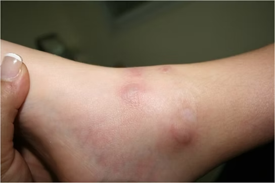
Other organ manifestations are frequent. Liver involvement is common; biopsy may show granulomas, though liver function is seldom significantly affected⁵. Cutaneous lesions occur in >25% (plaques, nodules, erythema nodosum, lupus pernio)⁵. Lupus pernio—often chronic—affects the face (nose, cheeks, lips, ears) and is associated with more severe disease⁵.
Associated Comorbidities
Articular disease (sarcoid arthropathy) affects 6–35%⁶, typically a bilateral polyarthritis of knees and ankles that is often self-limited over weeks to months⁶. Symptoms can be chronic or recurrent; joint destruction is rare⁶. Erythema nodosum and hilar lymphadenopathy may co-occur with arthritis—this triad is Löfgren’s syndrome, also generally self-limited⁶.
Muscle involvement can present as sarcoid myositis. Neurologic manifestations include mononeuropathy, polyneuropathy, and less commonly facial nerve palsy; polyneuropathy is usually symmetric but rare⁶.
Pharmacologic Treatment
Corticosteroids are first-line (prednisone/prednisolone)⁷.Antimalarials (chloroquine or hydroxychloroquine) are used for cutaneous disease or hypercalcemia⁷.For chronic disease, methotrexate can be steroid-sparing (benefit often after months)⁷.Azathioprine is an alternative but may cause nausea and neutropenia⁷.Other agents such as cyclophosphamide are rarely used due to toxicity⁷. Limited and weak evidence supports pentoxifylline, cyclosporine, infliximab⁷.
Diagnosis
Diagnosis rests on clinical assessment, history, and targeted testing⁸:
Chest radiograph (PA view) is the first step when pulmonary sarcoidosis is suspected—classically showing bilateral hilar lymphadenopathy⁸.
Pulmonary function tests (spirometry, lung volumes) assess ventilatory status⁸.
Laboratory tests (CBC, serum calcium, liver enzymes, creatinine, BUN) help uncover organ involvement⁸.
Biopsy of involved tissue (lymph node, skin, salivary gland) can confirm non-caseating granulomatous inflammation⁸.
Additional studies: urinalysis, ECG, ophthalmologic exam, and tuberculin testing to exclude mimics⁸.Key differentials: tuberculosis, lymphoma, carcinoma, berylliosis, and certain fungal infections⁸.Diagnosis is often delayed—especially for CNS and pulmonary disease—while cutaneous disease is typically recognized earlier⁸.
Etiology / Causes
No single definitive cause is known¹. The leading hypothesis is an immune response to an unknown trigger in genetically predisposed individuals². Familial and ethnic clustering supports genetic contributions². Potential triggers include viral or bacterial infections, dust particles, or chemical exposures². The broad clinical variability suggests sarcoidosis likely results from multiple factors that vary by person³.
Medical Management
Many patients have mild disease and do not require therapy, as spontaneous remission is common—especially in Stage I–II⁴. When symptoms are significant or organ function is compromised, corticosteroids are first-line⁵.
Prednisone is most commonly used to suppress inflammation and limit fibrosis⁵.
Dosing and duration vary; there is no universal protocol. Treatment is tailored to site, severity, and progression, with attention to minimizing adverse effects⁵.
If steroids are inadequate or poorly tolerated, consider methotrexate or azathioprine⁵.
For severe cutaneous disease or hypercalcemia, hydroxychloroquine may be beneficial⁵.
Cyclophosphamide and other potent immunosuppressants are reserved for severe, refractory cases due to toxicity⁵.
Physiotherapeutic Management
Physiotherapy should be individualized to symptoms and stage⁶.
In acute sarcoidosis, spontaneous regression is common, so physiotherapy may be minimal or unnecessary⁶.
In chronic disease or with substantial organ involvement—particularly lungs—physiotherapy aims to optimize lung function, increase ventilation, and improve physical capacity⁶.
Interventions may include:
Breathing exercises to improve ventilation and gas exchange.
Strength and endurance training to reduce dyspnea and increase work capacity⁶.
Combined upper- and lower-body exercise with respiratory training for maximal benefit⁶.
Goals: reduce functional limitations, increase stamina, lessen fatigue, and maximize daily function⁶.
Differential Diagnosis
Because sarcoidosis mimics many conditions, careful differential diagnosis is essential⁷.
Pulmonary mimics:
Tuberculosis
Atypical mycobacterial infection
Cryptococcosis
Aspergillosis
Histoplasmosis
Coccidioidomycosis
Blastomycosis
Hypersensitivity pneumonitis
Pneumoconiosis (beryllium)
Drug reactions
Aspiration
Granulomatosis with polyangiitis
Chronic interstitial pneumonia
Necrotizing sarcoid granulomatosis
Lymphatic diseases:
Atypical mycobacterial infection
Brucellosis
Toxoplasmosis
Granulomatous histiocytic necrotizing lymphadenitis
Cat-scratch disease
Reaction to carcinoma
Hodgkin disease
Non-Hodgkin lymphoma
GLUS syndrome
Cutaneous diseases:
Atypical mycobacterial infection
Fungal infections
Foreign-body reactions
Rheumatoid nodules
Hepatic diseases:
Brucellosis
Schistosomiasis
Primary biliary cholangitis
Crohn disease
Hodgkin disease
Non-Hodgkin lymphoma
GLUS syndrome
Bone marrow diseases:
Histoplasmosis
Infectious mononucleosis
Cytomegalovirus
Hodgkin disease
Non-Hodgkin lymphoma
Drug-related reactions
GLUS syndrome
Other:
Brucellosis
Various infections
Crohn disease
Giant cell myocarditis
GLUS syndrome
A thorough history, physical exam, and targeted laboratory testing are critical to distinguish sarcoidosis from these entities⁷.
References
Fuller KS, Goodman CC. Pathology implications for the physical therapist. 3rd ed. St. Louis: Saunders, 2009.
American Lung Association. Lung Diseases: sarcoidosis. http://www.lungusa.org/lung-disease/sarcoidosis/
Wu JJ, Schiff KR. Sarcoidosis. American Family Physician. 2004; 70,2: 312-322.
Ho LP, Urban BC, Thickett DR, Davies RJ. Deficiency of a subset of T cells with immunoregulatory properties in sarcoidosis. The Lancet. 2005; 365: 1062-1072.
Sève, P., Pacheco, Y., Durupt, F., Jamilloux, Y., Gerfaud-Valentin, M., Isaac, S., Boussel, L., Calender, A., Androdias, G., Valeyre, D., & Jammal, T. E. (2021). Sarcoidosis: A Clinical Overview from Symptoms to Diagnosis. Cells, 10(4), 766.
Gjennomgått - Trukket
Medicine Net. Sarcoidosis.http://www.medicinenet.com/sarcoidosis/page6.htm
MD Guidelines. Sarcoidosis. http://www.mdguidelines.com/sarcoidosis


