

Thoracic Hyperkyphosis
- Fysiobasen

- Dec 24, 2025
- 8 min read
Thoracic hyperkyphosis is defined as an increased forward curvature of the thoracic spine greater than 40°¹. Prevalence rises with age, but there is no single universally accepted definition of hyperkyphosis or of “normal” age-related curvature². It is more common in women than in men.
Normal thoracic curves:
20–40° in youth
48–50° in older women, ~44° in older men²
Subtypes
Scheuermann’s disease³: Juvenile form due to vertebral growth disturbance; typically halts at skeletal maturity.
Postural kyphosis³: Most common; no vertebral deformity. Related to poor posture, weak musculature, and ligament laxity.
Congenital kyphosis³: Present at birth; progresses without treatment.
Age-related hyperkyphosis⁴⁵: Common in older adults. Many have normal bone density. Estimated prevalence in those >50 years is 10–45%. In those >65 years: ~50% of men and ~65% of women⁶.
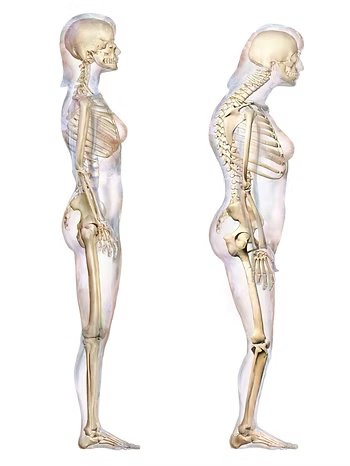
Anatomy
The thoracic spine normally has 20–40° of kyphosis¹⁷ in the sagittal plane. Hyperkyphosis exceeds 40°⁸. In standing, the line of gravity shifts anterior to the vertebral bodies, increasing loading and potentially amplifying kyphosis. Typical changes include tight posterior ligaments and increased tone/shortening of deep spinal extensors and long back extensors⁹.
Epidemiology & Causes
Rising prevalence with age, especially in women after 50.
Estimated prevalence: 20–40% in people over 60⁶.
Kyphosis angle increases ~9° per decade⁶.
Risk factors: musculoskeletal and neuromuscular disorders, sensory deficits, idiopathic causes⁵¹¹¹².
Psychosocial factors: depression, anxiety, insecurity, distress¹¹³.
Biomechanical Contributors
Increased spinal load and muscular demand in upright posture accelerate degeneration and contribute to pain¹³.
Habitual posture (forward head, limited shoulder mobility) from poor ergonomics and heavy schoolbags can worsen curvature⁴¹⁴.
Clinical Presentation
Thoracic hyperkyphosis may be postural (flexible) or structural (fixed); many patients have elements of both.
Postural kyphosis: Reversible with cueing and correction⁷.
Structural kyphosis: Permanent deformity (e.g., Scheuermann’s). Postural kyphosis can become structural over time⁷.
Common findings:
Visible “rounded back”⁵
Gradual onset/progression⁵¹⁵¹⁶
Thoracic back pain
Reduced pulmonary function⁹¹⁷
Decreased mobility
Osteoporotic vertebral fractures¹⁸
Difficulty rising, walking, or balancing
Fatigue
Dyspnea in severe deformity¹⁵
Differential Diagnosis
Scheuermann’s disease
Osteoporosis
Traumatic changes (e.g., compression fracture)
Tumor
Infection
Degenerative disc disease⁵¹⁹
Diagnostic Work-up
Radiography
First-line imaging is conventional X-ray. Obtain both AP and lateral views²⁰²¹.
AP view: Assesses vertebral bodies.
Lateral view: Assesses vertebral height, disc height, end-plate irregularities, erosions, and curve alignment²⁰.
If needed, CT or MRI can further delineate spinal curvature and characterize kyphosis²¹.
Outcome Measures
Occiput-to-wall distance: Forward head/trunk measure.
VAS (Visual Analogue Scale): Pain intensity²².
Quebec Back Pain Disability Scale: Self-report functional status²³.
Examination
Typical age-related hyperkyphosis features:
Pain and dysfunction in back and shoulders¹³
Reduced ROM and increased stiffness
Limited physical function²⁴
Respiratory difficulty
Increased risk of osteoporotic fractures²⁵
Increased mortality in older adults
Begin with observation of gait and movement (e.g., while undressing)¹⁶. Assess standing posture in neutral (feet under hips, knees extended, arms relaxed, gaze horizontal)³¹⁶. Tragus-to-wall testing may be used. A thoracic Cobb angle of 40–45° indicates hyperkyphosis.
Tools to Quantify Kyphosis
Modified Cobb angle
Pantograph
Debrunner kyphometer: Landmarks at C7 and T12; read angle on dial.
Flexicurve: Mold from C7 to lumbosacral junction; trace on graph paper for index calculations.
Medical Management
Physical (first-line): Conservative physical care is recommended initially for thoracic hyperkyphosis⁵.
Pharmacologic: Antiresorptive or anabolic bone medications in low bone mass or vertebral fractures can prevent new fractures but have not shown direct reduction of hyperkyphosis itself⁵.
Surgical: Approach depends on flexibility:
Flexible curves: Posterior approach; otherwise anterior or combined approach with instrumented fusion²⁶.
Osteotomy: Selected cases may benefit.
Osteoporotic collapse: Kyphoplasty may restore height with balloon and cement.
Vertebroplasty/Kyphoplasty: Minimally invasive options with evidence for pain relief, functional improvement, height restoration (up to ~90%), and kyphosis correction of ~8.5–14°²⁶ (overall evidence still limited²²⁶).
Extensive surgery carries substantial risk (~33% complications) and is reserved for documented progression, severe pain, or neurologic deficits⁵.
Physiotherapy for Thoracic Hyperkyphosis

Early, structured physiotherapy—manual therapy, taping, bracing, and individualized exercise—can reduce curvature, improve posture, and slow progression. Goals include better biomechanics and function to reduce pain, enhance mobility, and prevent complications such as reduced ventilation and balance deficits²⁷.
Treatment goals:
Reduce excessive thoracic kyphosis angle
Improve postural control and alignment
Increase joint and soft-tissue mobility in the thoracic spine
Prevent progression and sequelae
Decrease back/shoulder pain
Strengthen spinal and core stabilizers
Improve lung capacity and breathing mechanics
Promote participation in ADLs and quality of life⁵
Manual Therapy
Thoracic mobilization
Facet-focused mobilization to improve flexibility and reduce stiffness.
Myofascial techniques for tight musculature and fascia⁵.
Scapular mobilization to normalize shoulder mechanics commonly altered in hyperkyphosis.
Self-mobilization
Diaphragmatic breathing lying on a foam roller to encourage thoracic expansion and better breathing mechanics.
Thoroughly instructed home drills to maintain mobility⁵.
Stretching
Targeted flexibility
Pectoralis major/minor stretch (e.g., on foam roller)
Prone hip-flexor stretch (iliopsoas/rectus femoris)
Supine hamstring stretch with 90° hip flexion²⁸
Corrective Exercise
Postural training
Scapular stabilization: Strengthen mid-lower trapezius, rhomboids, serratus to support alignment.
Spinal extensor training: Counteract excessive kyphosis and improve endurance.
Aim to enhance postural control, reduce stiffness, and foster structural adaptation²⁹³⁰.
Pain Modulation
Heat, cold, and TENS may aid short-term pain relief, especially in acute flares⁵.
Strengthening
Examples
Prone trunk lifts to neutral: strengthens extensors and trapezius.
Trunk lifts with backpack load: progressive extensor loading.
Quadruped contralateral arm/leg raise: improves stability and reduces forward collapse⁵.
Breathing Training
Diaphragmatic breathing
Improves oxygenation and activity tolerance, increases rib mobility, and reduces dyspnea.
Balance & Gait Training
Integrate balance and gait work to lower fall risk (often elevated with hyperkyphosis). Can be incorporated via Pilates-style or tailored stabilization programs.
Yoga
A safe adjunct to blend strength, mobility, breath, and relaxation; RCTs show benefit in posture and function²⁹³¹³².
Bracing
Indications
Consider in stiff curves or when exercise alone is insufficient. Always pair with physiotherapy; bracing alone has limited effect²⁷⁵¹⁶.
Common braces
Milwaukee brace: Posterior pads apply corrective force; typically 23 h/day for 1–2 years¹⁶.
Lyon Antikyphosis Brace: Effective in many adolescents/adults²⁷.
Kyphologic Brace: Modern design with documented in-brace correction³³.
Gschwend brace: Widely used in parts of Europe²⁸.
Taping
From AC joint anteriorly, over trapezius, diagonally to ~T6 may reduce kyphosis; more research needed⁵.
Spinal Orthosis
SpinoMed
~2 h/day for 6 months can reduce kyphosis angle, increase standing height, strengthen extensors, and reduce instability⁵.
Conclusion
International evidence supports conservative care—systematic physiotherapy with stretching, strengthening, manual therapy, balance and breathing work, plus judicious use of bracing and activity modification—as effective for thoracic hyperkyphosis²⁸. Individualized programs yield the best outcomes.
References:
Fon GT, Pitt MJ, Thies Jr AC. Thoracic kyphosis: range in normal subjects. American Journal of Roentgenology. 1980 May 1;134(5):979-83.
Kado DM, Prenovost K, Crandall C. Narrative review: hyperkyphosis in older persons. Annals of internal medicine. 2007 Sep 4;147(5):330-8.
de Mauroy JC. Kyphosis physiotherapy from childhood to old age. InPhysical Therapy Perspectives in the 21st Century-Challenges and Possibilities 2012 Apr 5. IntechOpen.
Perriman DM, Scarvell JM, Hughes AR, Lueck CJ, Dear KB, Smith PN. Thoracic hyperkyphosis: a survey of Australian physiotherapists. Physiotherapy Research International. 2012 Sep;17(3):167-78.
Katzman WB, Wanek L, Shepherd JA, Sellmeyer DE. Age-related hyperkyphosis: its causes, consequences, and management. journal of orthopaedic & sports physical therapy. 2010 Jun;40(6):352-60.
Bartynski WS, Heller MT, Grahovac SZ, Rothfus WE, Kurs-Lasky M. Severe thoracic kyphosis in the older patient in the absence of vertebral fracture: association of extreme curve with age. American journal of neuroradiology. 2005 Sep 1;26(8):2077-85.
Sahrmann S. Movement system impairment syndromes of the extremities, cervical and thoracic spines-e-book. Elsevier Health Sciences; 2010 Nov 19.
Ackland TR, Elliott B, Bloomfield J. Applied anatomy and biomechanics in sport. Human Kinetics; 2009.
Edmondston SJ, Singer KP. Thoracic spine: anatomical and biomechanical considerations for manual therapy. Manual therapy. 1997 Aug 1;2(3):132-43.
Nishiwaki Y, Kikuchi Y, Araya K, Okamoto M, Miyaguchi S, Yoshioka N, Shimada N, Nakashima H, Uemura T, Omae K, Takebayashi T. Association of thoracic kyphosis with subjective poor health, functional activity and blood pressure in the community-dwelling elderly. Environmental health and preventive medicine. 2007;12(6):246-50.
↑ Negrini S, Aulisa AG, Aulisa L, Circo AB, de Mauroy JC, Durmala J, Grivas TB, Knott P, Kotwicki T, Maruyama T, Minozzi S. 2011 SOSORT guidelines: orthopaedic and rehabilitation treatment of idiopathic scoliosis during growth. Scoliosis. 2012 Dec;7(1):3.
Ashton-Miller JA. Thoracic hyperkyphosis in the young athlete: a review of the biomechanical issues. Current sports medicine reports. 2004 Jan 1;3(1):47-52.
Lewis JS, Valentine RE. Clinical measurement of the thoracic kyphosis. A study of the intra-rater reliability in subjects with and without shoulder pain. BMC musculoskeletal disorders. 2010 Dec;11(1):39.
Britnell SJ, Cole JV, Isherwood L, Stan MM, Britnell N, Burgi S, Candido G, Watson L. Postural health in women: the role of physiotherapy. Journal of obstetrics and gynaecology Canada. 2005 May 1;27(5):493-500.
Zane MK. Physical Therapist’s Guide to Hyperkyphosis (Humpback) in Adults. American Physical Therapy Association. 2014;188.
Zaina F, Atanasio S, Ferraro C, Fusco C, Negrini A, Romano M, Negrini S. Review of rehabilitation and orthopedic conservative approach to sagittal plane diseases during growth: hyperkyphosis, junctional kyphosis, and Scheuermann disease. Eur J Phys Rehabil Med. 2009 Dec 1;45(4):595-603.
Culham EG, Jimenez HA, King CE. Thoracic kyphosis, rib mobility, and lung volumes in normal women and women with osteoporosis. Spine. 1994 Jun;19(11):1250-5.
Greendale GA, Nili NS, Huang MH, Seeger L, Karlamangla AS. The reliability and validity of three non-radiological measures of thoracic kyphosis and their relations to the standing radiological Cobb angle. Osteoporosis international. 2011 Jun 1;22(6):1897-905.
D'Antoni AV, Terzulli SL. Federico di Montefeltro's hyperkyphosis: a visual-historical case report. Journal of medical case reports. 2008 Dec;2(1):11.
El-Khoury GY, Whitten CG. Trauma to the upper thoracic spine: anatomy, biomechanics, and unique imaging features. AJR. American journal of roentgenology. 1993 Jan;160(1):95-102.
Naidich TP, Castillo M, Cha S, Raybaud C, Smirniotopoulos JG, Kollias S. Imaging of the Spine: Expert Radiology Series, Expert Consult-Online and Print. Elsevier Health Sciences; 2010 Aug 27.
Jensen MP, Chen C, Brugger AM. Interpretation of visual analog scale ratings and change scores: a reanalysis of two clinical trials of postoperative pain. The Journal of pain. 2003 Sep 1;4(7):407-14.
Kopec JA, Esdaile JM, Abrahamowicz M, Abenhaim L, Wood-Dauphinee S, Lamping DL, Williams JI. The Quebec Back Pain Disability Scale. Measurement properties. Spine. 1995 Feb;20(3):341-52.
Kado DM, Huang MH, Barrett-Connor E, Greendale GA. Hyperkyphotic posture and poor physical functional ability in older community-dwelling men and women: the Rancho Bernardo study. The Journals of Gerontology Series A: Biological Sciences and Medical Sciences. 2005 May 1;60(5):633-7.
Huang MH, Barrett‐Connor E, Greendale GA, Kado DM. Hyperkyphotic posture and risk of future osteoporotic fractures: the Rancho Bernardo study. Journal of Bone and Mineral Research. 2006 Mar;21(3):419-23.
Lundine K, Turner P, Johnson M. Thoracic hyperkyphosis: assessment of the distal fusion level. Global spine journal. 2012 Jun;2(02):065-70.
de Mauroy JC, Vallèse P, Fender P, Lecante C. Historical Lyonaise brace treatment for adolescent hyperkyphosis. Results of 272 cases reviewed 2 years minimum after removal of the brace. Scoliosis. 2010 Sep;5(1):O69.
Weiss H, Turnbull D. Kyphosis (Physical and technical rehabilitation of patients with Scheuermann's disease and kyphosis). International encyclopedia of rehabilitation. 2010.
Jang HJ, Kim MJ, Kim SY. Effect of thorax correction exercises on flexed posture and chest function in older women with age-related hyperkyphosis. Journal of physical therapy science. 2015;27(4):1161-4.
Wang MY, Greendale GA, Kazadi L, Salem GJ. Yoga improves upper-extremity function and scapular posturing in persons with hyperkyphosis. Journal of yoga & physical therapy. 2012 Jun 1;2(3):117.
Greendale GA, Huang MH, Karlamangla AS, Seeger L, Crawford S. Yoga decreases kyphosis in senior women and men with adult‐onset hyperkyphosis: results of a randomized controlled trial. Journal of the American Geriatrics Society. 2009 Sep;57(9):1569-79.
Kuo YL, Tully EA, Galea MP. Sagittal spinal posture after Pilates-based exercise in healthy older adults. Spine. 2009 May 1;34(10):1046-51.
Weiss HR, Werkmann M, Bohr S. In-brace corrections in patients with kyphosis using the kyphologic® brace. Scoliosis. 2009 Dec 1;4(S2):O61.


