

Achilles Tendon Rupture

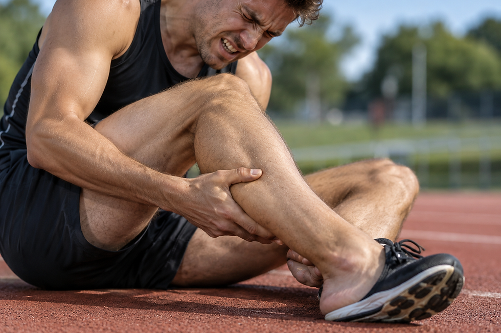
The Achilles tendon is a strong band of connective tissue that connects the calf muscles to the heel bone (calcaneus).
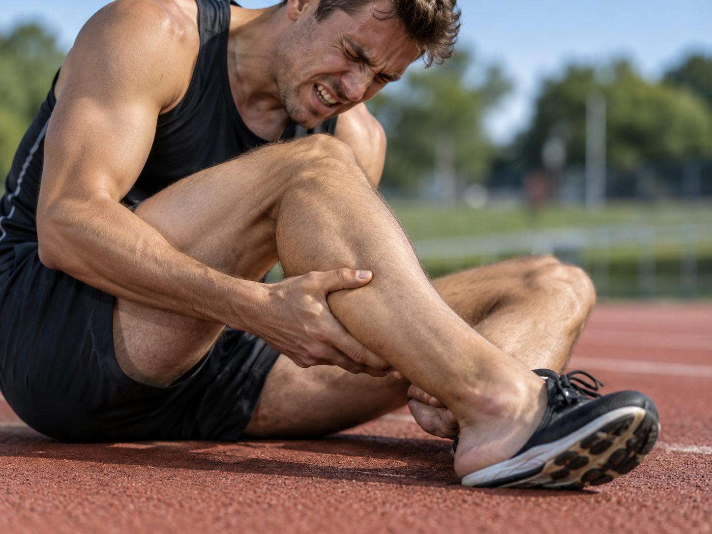
Achilles tendon rupture (ATR) is a common injury and is considered the most frequently ruptured tendon in the human body.[1][2] The condition can have a significant negative impact on both physical and mental health.[3][4] The main causes of rupture include:
Overstretching of the heel during sports activities.
Forceful plantar flexion of the foot.
Falls from height.[5]
Clinically Relevant Anatomy
Anatomy of the Calf Muscles
The Achilles Tendon:
The longest and strongest tendon in the human body, consisting of strong and relatively inelastic connective tissue.[6]
Serves as the attachment of the soleus and gastrocnemius muscles to the calcaneal tuberosity (tuber calcanei).
The calf muscles are primarily responsible for plantar flexion of the ankle.
Epidemiology
The incidence of Achilles tendon ruptures has increased in recent years, which is associated with higher rates of obesity and increased participation in recreational sports.[7][8]
The incidence is 7 per 100,000 per year in the general population, but rises to 15–25 per 100,000 per year among recreational athletes.[9][10]
The injury occurs most frequently in the 30–50-year age group.
It typically affects "middle-aged weekend warriors," although this group accounts for only about 70% of cases.
Men are affected significantly more often than women.[11]
Etiology
Because of its intrinsic properties, function, and blood supply, the Achilles tendon is predisposed to both acute and chronic ruptures.
Acute Ruptures
Acute ruptures often occur during sporting activities and account for approximately 68% of injuries.[11][12]
The injury is common in sports such as badminton, football, volleyball, basketball, tennis, squash, and other stop-and-go sports in which eccentric movements place high loads on the tendon.
Factors that predispose to Achilles tendon injuries include:
Natural aging: Reduced tissue elasticity and strength.[12]
Recreational sports: Sudden loading of the tendon.
Obesity: Increased mechanical load.[12]
Medications: Use of fluoroquinolones and steroids is associated with increased risk.
Biomechanical abnormalities: Such as flat feet (pes planus), high arches (pes cavus), or leg-length discrepancy.
Poor running technique: Results in suboptimal loading of the tendon.
Characteristics and Clinical Presentation
Mechanisms of Injury
Three main types of indirect injury can lead to Achilles tendon rupture:[5]
Push-off with the weight on the forefoot while the knee is extending, typically during sprinting, running, or jumping.
Sudden and unexpected dorsiflexion of the ankle, for example when slipping off a chair, tripping, or falling forward.
Forceful dorsiflexion of a plantar-flexed foot, such as in a fall from height.
Acute Rupture: Signs and Symptoms
The patient experiences sharp pain, often described as being "stabbed in the heel with a knife."[13][14][15][1][16][9]
The injury is often accompanied by an audible snap or popping sound.[13][16]
On palpation, a palpable gap in the tendon may be felt.[13][14][15]
Visible signs include:
Swelling at the back of the heel.
Reduced active plantar flexion of the ankle.[9]
Increased passive dorsiflexion.
Inability to lift the heel off the ground.
Difficulty walking.
Thompson test: A positive test often confirms the diagnosis.
Some patients may have a history of tendinopathy or previous corticosteroid injections in the area.[13][14][15]
Chronic Rupture
Ruptures are considered chronic if the injury is identified 4–6 weeks after the time of injury.[5]
Symptoms of chronic rupture include:
Pain, particularly during activity.
Reduced strength in the calf and foot musculature.
Fatigue and stiffness in the ankle.[17]
An accurate diagnosis is essential because management differs from that of acute cases.
This comprehensive overview provides a basis for differential diagnosis and appropriate management of Achilles tendon injuries, whether acute or chronic.
Differential Diagnosis
When assessing an Achilles tendon injury, other conditions should be excluded to ensure an accurate diagnosis. Possible differential diagnoses include:
Acute Achilles tendon inflammation (peritendinitis)
Rupture or injury of the gastrocnemius muscle
Strain or rupture of the calf musculature
Posterior tibialis muscle stress syndrome
Fracture of the ankle or nearby structures
Posterior tibialis tendon injuries
Peroneal tendon injuries
Classification of Achilles Tendon Ruptures
Achilles tendon injuries are traditionally classified into four types based on injury severity and the degree of tendon retraction. This classification has traditionally guided treatment decisions:
Type I: Partial rupture ≤50%
Usually treated conservatively with immobilization and physiotherapy.
Type II: Complete rupture with a tendon gap ≤3 cm
Typically treated with end-to-end suturing (anastomosis).
Type III: Complete rupture with a tendon gap of 3–6 cm
Often requires the use of a tendon graft or synthetic graft.
Type IV: Complete rupture with a tendon gap >6 cm (neglected ruptures)
Often treated with a tendon or synthetic graft combined with gastrocnemius lengthening.
Newer Insights Into Treatment Selection
Helene Simpson[19] highlights that newer research questions basing treatment decisions solely on the size of the rupture or gap. According to Simpson, an injured Achilles tendon should be treated as if 100% of the tendon fibers are affected, regardless of the actual gap size or the proportion of fibers involved.
The choice of treatment, whether surgical or conservative, should be guided by a range of factors, including:
The patient's medical history
Their activity level and functional demands
Financial circumstances and available treatment options
Degree of functional loss and expected rehabilitation course
This holistic approach ensures that treatment is tailored to the individual patient and takes both medical and social factors into account.
Examination When Achilles Tendon Rupture Is Suspected
Observation
When inspecting a suspected Achilles tendon rupture, the therapist can observe the patient in different positions to identify signs of injury:
Standing
Assess for signs of collapsed arches (flat feet) and other postural abnormalities.
Prone
The patient is usually positioned prone. This allows closer observation of the tendon for signs of:
Thickening
Redness
Swelling
Nodular changes
Walking and Running
Observe for overpronation and other gait abnormalities.
Swelling
A swollen ankle may indicate an Achilles tendon rupture.
Palpation
The Achilles tendon is easy to palpate. Palpation along the entire length of the tendon may reveal a gap, which is a classic sign of rupture.
Active Movements
In this part of the examination, the patient is instructed to perform active movements that may help diagnose a tendon injury.
Typical active movements include:
Observation of gait pattern
With an Achilles tendon rupture, the patient may have difficulty performing plantar flexion at the ankle. Signs of a possible rupture include:
Painful and inhibited plantar flexion during gait.
Overpronation of the injured ankle.
Loss of push-off at the end of stance phase due to dysfunction of the gastrocnemius and soleus muscles.
Standing on Tiptoes
The patient is instructed to stand on tiptoes to perform plantar flexion. This will be impossible with an Achilles tendon rupture.
Active Plantar Flexion
The patient is asked to actively perform plantar flexion at the ankle.
Any movement involving plantar flexion will be severely limited or impossible with a complete rupture.
These examinations provide a comprehensive assessment of functional status and important information for confirming the diagnosis and planning further management.
Special Tests for Achilles Tendon Rupture
Several special tests can be used to diagnose Achilles tendon rupture and assess its extent:
1. Thompson Test
This test is particularly useful for diagnosing complete Achilles tendon ruptures, but is less suitable for detecting partial ruptures.
Procedure: The patient lies prone, and the therapist squeezes the patient's calf muscles.
Result: A negative test produces plantar flexion of the foot when the calf muscles are squeezed. In a positive test, plantar flexion is absent, indicating a complete rupture.
2. Matles Test
This test is considered the most reliable for Achilles tendon rupture, particularly in the emergency department.
Procedure: The patient lies prone and is asked to actively flex the knee to 90 degrees. The therapist observes the movement of the foot and ankle.
Result:
Negative test: The foot shows slight plantar flexion.
Positive test: The foot falls into a neutral position or shows dorsiflexion during the movement.
Sensitivity: Reported sensitivity is 0.88¹.
3. ATRS (Achilles Tendon Total Rupture Score)
This is a questionnaire that assesses the limitations and challenges experienced by a patient with an Achilles tendon rupture.
It is important for evaluating the patient's functional status and perceived treatment outcomes.
4. Ultrasound-Assisted Thompson Test
This test combines the classic Thompson test with real-time ultrasound visualization.
Procedure: The calf muscle is squeezed while ultrasound is used to assess the response in the Achilles tendon.
Use: It can be performed by surgeons with minimal ultrasound training and provides improved diagnostic accuracy compared with static ultrasound.
Reasons Diagnostic Assessment May Fail
Achilles tendon rupture is often missed in the emergency department because:
The squeeze test and static ultrasound are not always reliable.
The Matles test is considered the most accurate diagnostic method¹.
Outcome Measures
Objective Measures
These parameters are recorded directly by the physiotherapist and provide measurable data on the patient's physical function:
Ankle range of motion (ROM): Assesses the degree of plantar flexion and dorsiflexion.
Calf muscle strength: Tests the strength of the gastrocnemius and soleus muscles.
Patient-Reported Measures
Over the past two decades, it has become clear that the patient's own assessment of outcomes is essential for evaluating treatment success.
ATRS (Achilles Tendon Rupture Score): A condition-specific questionnaire that evaluates the patient's functional limitations.
SF-36 (Short Form-36): A generic questionnaire that measures health-related quality of life. It is a valid and reliable tool used to monitor health status and treatment effects.
Recommendations for Outcome Measures
To obtain a comprehensive picture of treatment outcomes, the following should be combined:
Patient-reported outcomes: Focus on the patient's perception of health status and quality of life.
Objective indicators:
Muscle strength
Calf muscle endurance
Ability to return to the previous activity level
Such an approach provides a more complete understanding of treatment effectiveness and the patient's functional status.
Chronic Achilles Tendon Rupture
When assessing an Achilles tendon rupture that has been present for a longer period, the presence of tendon stumps and the length of the rupture gap play a decisive role in the choice of surgical treatment. Several surgical options may be used, including:
V-Y lengthening
Gastrocnemius fascial turndown flap
Plantaris tendon harvesting
Achilles tendon scraping
Flexor hallucis longus (FHL) tendon transfer
These techniques aim to restore tendon function and strength, especially in complex or neglected cases.
Acute Achilles Tendon Rupture
Traditionally, open surgical repair has been used to treat acute rupture, but this method has a high complication rate, including:
Wound infections
Abnormal sensation
Adhesions
Thrombosis
To reduce these complications, newer techniques using smaller incisions have been developed. Minimally invasive techniques and percutaneous repair are becoming increasingly popular because they reduce complications while providing good outcomes.
Complications After Surgery
Sural nerve entrapment: Common during suturing, particularly in endoscopic procedures.
Tibial nerve: May also become involved, leading to pain.
Symptoms: Patients often report pain on the lateral side of the ankle early in the rehabilitation phase.
Neurological assessment: Important for identifying the source of pain and avoiding further complications. Although the pain often decreases over time, it should be monitored carefully.
Treatment
Research shows that conservative treatment can provide good outcomes after Achilles tendon rupture. Several studies have shown that tendon strength after conservative treatment is comparable with surgical treatment, while the risk of rerupture is low, provided the rupture is identified within 48 hours of injury.
The Importance of Early Rehabilitation
Between 2011 and 2017, several meta-analyses were published examining the effect of conservative treatment for Achilles tendon rupture. The main finding was that early rehabilitation and mobilization are key to good outcomes. Compared with surgical treatment, it is the introduction of early rehabilitation that distinguishes a successful conservative approach from less effective methods.
Goals of Conservative Treatment
The main goal is to restore and maintain contact between the two ends of the ruptured Achilles tendon to facilitate the healing process.
Typical Conservative Treatment Protocol

Immobilization with a rigid cast or functional orthosis.
Foot position:
Begin in full equinus (30° plantar flexion).
Gradually bring the foot to a neutral position over a period of 8–12 weeks.
Weight-bearing is allowed as soon as the ankle position permits.
There is no clinical consensus on whether the cast should extend above the knee or whether a below-knee cast is sufficient.
This approach allows the patient to achieve functional recovery without surgery while reducing the risk of complications.
Chronic Achilles Tendon Rupture
In chronic Achilles tendon rupture, the presence or absence of tendon stumps and the length of the rupture gap are assessed to select the appropriate surgical technique. The following surgical options may be considered:
V-Y lengthening
Gastrocnemius fascial turndown flap
Plantaris tendon harvesting
Achilles tendon scraping
Flexor hallucis longus (FHL) tendon transfer
These techniques are tailored to repair the tendon and restore function, especially in complex or neglected cases.
Acute Achilles Tendon Rupture
Open surgical repair for acute rupture has traditionally been used but has a high complication rate, including:
Wound infection
Abnormal sensation
Adhesions
Thrombosis
To reduce these complications, newer techniques with smaller incisions have been introduced. Minimally invasive techniques and percutaneous repair are becoming increasingly popular because they reduce complications and provide good outcomes.
Complications After Surgery
Sural nerve entrapment: May occur during suturing, particularly in endoscopic procedures.
Tibial nerve: May be involved and cause pain in the postoperative phase.
Symptoms: Patients often report pain on the lateral side of the ankle early in the rehabilitation phase.
A thorough neurological assessment is important for identifying the cause of pain and preventing further complications. Although the pain often decreases over time, it should be monitored to ensure appropriate rehabilitation.
Conservative Treatment
Research shows that conservative treatment can provide good outcomes for patients with Achilles tendon rupture, especially if the injury is identified early, within 48 hours of injury.
Studies have shown that tendon strength after conservative treatment is comparable with surgical treatment and that the rerupture rate is low.
The Importance of Early Rehabilitation
Between 2011 and 2017, several meta-analyses were published examining conservative treatment for Achilles tendon rupture. These showed that the key to success is early mobilization and rehabilitation. When surgical treatment is compared with conservative methods, inclusion of early rehabilitation is what accounts for the difference in outcomes.
Goals of Conservative Treatment
The main goal is to restore and maintain contact between the two ends of the ruptured tendon to facilitate healing.
Typical Conservative Treatment Protocol
Immobilization: Usually with a rigid cast or functional orthosis.
Foot position:
Begin in full equinus (30° plantar flexion).
Gradually adjust to a neutral position over 8–12 weeks.
Weight-bearing: Allowed as soon as the ankle position permits.
There remains uncertainty about whether the cast should extend above the knee or whether a below-knee cast is sufficient.
This approach promotes healing while avoiding complications such as rerupture and impaired function.
Conservative Treatment: Accelerated Functional Rehabilitation Approach
Principles of Early Mobilization
Regardless of the size or extent of the rupture, conservative treatment may be recommended if the patient demonstrates a high level of adherence and follows an early mobilization program closely[19].
Although support is increasing for accelerated rehabilitation regimens after both surgical and nonsurgical treatment, there is still no consensus regarding the optimal protocol, timing of initiation, or types of exercises that should be included[33].
The First Two Weeks
Immobilization:
The patient should avoid all dorsiflexion and keep the foot immobilized in an orthosis or cast in 30° plantar flexion.
Avoid weight-bearing:
Weight-bearing is discouraged during this period to protect the tendon.
Early postoperative measures:
In selected cases, depending on the surgical procedure and condition, the patient may begin gradual weight-bearing under supervision as early as 24 hours after surgery. This involves bearing approximately 5 kg of weight in a boot positioned at 30° plantar flexion while using a scale for feedback, always under medical supervision.
Preparation for physiotherapy:
The patient can then prepare for physiotherapy by using crutches and a boot for support and balance. Isometric quadriceps exercises are recommended to counteract muscle atrophy. In addition, ankle-pump exercises may be used to reduce edema, combined with 15 minutes of cold therapy every two hours.
Two to Four Weeks
After the first two weeks, the patient can begin simple, controlled mobilization exercises:
Briefly remove the boot:
The patient is encouraged to remove the orthosis for five minutes every hour or every other hour to perform specific exercises. The focus is on improving plantar-flexion mobility and gradually strengthening the muscles involved in this movement.
Avoid dorsiflexion:
Dorsiflexion stretching and movement toward neutral are still prohibited to protect the healing process.
Four to Eight Weeks
Gradual reduction of heel wedges:
Between weeks 4 and 8, the angle of the boot wedges is gradually reduced, with the goal of reaching a neutral position by week 10.
Adaptation to the patient's needs:
Adjustments to the protocol are based on the surgical technique, the patient's medical condition, and any comorbidities.
Areas of Focus
Early mobilization:
The aim is to promote tendon healing through cautious loading and controlled movement.
Individualized approach:
The patient's condition, medical history, and level of adherence influence rehabilitation progression.
This accelerated rehabilitation method combines structured immobilization with gradual mobilization and strengthening to achieve optimal function and reduce the risk of rerupture.
Conservative Treatment – Accelerated Functional Rehabilitation Approach
Principles of Early Mobilization
Regardless of the size and severity of the rupture, conservative treatment may be recommended if the patient demonstrates a high level of adherence and follows a strict early mobilization program[19].
Although support is increasing for accelerated rehabilitation regimens after both surgical and nonsurgical treatment, there is still no consensus on the optimal protocol, when rehabilitation should begin, or which exercises should be included[33].
The First Two Weeks
Immobilization:
The patient should avoid all dorsiflexion and immobilize the foot in 30° plantar flexion using an orthosis or cast.
Avoid weight-bearing:
Weight-bearing is recommended to be avoided during the first two weeks to protect the tendon and promote healing.
Gradual weight-bearing (selected cases):
In some cases, the patient may begin gradual weight-bearing as early as 24 hours after surgery, depending on the surgeon's assessment. This involves bearing approximately 5 kg of weight in a boot at 30° plantar flexion, under close medical supervision.
Preparation for physiotherapy:
After this period, the patient can be prepared for physiotherapy using crutches and a boot for support and balance. Isometric quadriceps exercises are recommended to maintain muscle strength, while ankle-pump exercises may help reduce edema. Cold therapy for 15 minutes every two hours is recommended to manage swelling.
Two to Four Weeks
Brief removal of the orthosis:
The patient is encouraged to remove the boot for five minutes every hour or every other hour to perform specific exercises.
Focus on plantar flexion:
Exercises focus on improving plantar-flexion mobility and gradually strengthening the muscles involved in this movement.
Avoid dorsiflexion:
Dorsiflexion and movement toward a neutral position are still not permitted in order to protect the healing process.
Four to Eight Weeks
Gradual lowering of heel wedges:
From weeks 4 to 8, the angle of the boot wedges is gradually reduced, with the goal of reaching neutral by week 10.
Individualized progression:
The protocol is adapted based on the surgical technique, the patient's medical history, and the presence of comorbidities.
Rehabilitation Focus Areas
Early mobilization:
Combines cautious loading with controlled movement to promote healing and functional improvement.
Individualized approach:
The rehabilitation regimen is adapted to the patient's specific needs, activity level, and medical condition.
This accelerated functional rehabilitation approach balances immobilization and gradual mobilization to ensure optimal healing, reduce the risk of rerupture, and
restore functional capacity.
Traditional Approach to Rehabilitation After Achilles Tendon Rupture
Initial Phase: Immobilization
Rehabilitation after an Achilles tendon rupture depends on the initial treatment strategy, whether surgical or nonsurgical. During the first weeks, the ankle is normally immobilized using a cast or orthosis (below-knee posterior splint) that allows limited plantar flexion to facilitate early mobilization.
After removal of the cast or orthosis, rehabilitation focuses on:
Gradual improvement of ankle range of motion (ROM).
Gradual increase in muscle strength and coordination.
Gradual return to activities, usually after 4–6 months of treatment.
Rehabilitation progression depends strongly on the quality of treatment and the patient's motivation.
Early Phase: General Exercises (During Immobilization)
Physiotherapy can begin immediately during immobilization to maintain general strength and function in the injured leg. Possible exercises include:
Hip abduction: To maintain hip function.
Straight leg raise: Focus on quadriceps activation.
Standing hamstring curl: For the posterior thigh musculature.
Core muscle training (plank): For general stability.
After Removal of Cast or Orthosis (2 Weeks)
Focus:
Passive mobilization: Begin with gentle passive mobilization of the ankle and subtalar joints.
Active ROM exercises: Exercises such as ankle circles (clockwise and counterclockwise) and active flexion/extension.
Resistance training: After two weeks, progression to resistance exercises may be introduced.
Possible Exercises:
Active ankle flexion and extension.
Circular ankle movements.
Exercises from the early phase continue in parallel, such as hip abduction and hamstring curls.
Cycling on a stationary bike to improve endurance and circulation.
8–12 Weeks After Surgery
Focus:
Gait training: Begin with exercises to improve gait and activity-specific movements.
Resistance exercises with a resistance band: Start with the lowest resistance and gradually increase it.
Previous exercises: Continue exercises from earlier phases.
Examples of Exercises:
Ankle movement in plantar flexion and dorsiflexion to 90°: 2 x 8 repetitions.
Inversion and eversion: Focus on control and stability.
Pronation and supination: 2 x 8 repetitions.
Single-leg balance exercise: 3 x 30 seconds.
Standing calf raises (2-second tempo): 3 x 10 repetitions.
12–24 Weeks After Surgery
Focus:
Progressive strength training: Use ankle weights to increase resistance in the exercises.
Advanced exercises: Introduce balance and coordination exercises on unstable surfaces.
New Exercises:
Calf stretching.
Calf raises with increased load on the injured leg: 5 x 10 repetitions.
Balance exercises on a trampoline: 2 x 45 seconds.
Walking/running on a trampoline: 2 x 45 seconds.
Squats with the injured leg in front: 2 x 10 repetitions.
Single-leg press: 2 x 10 repetitions (maximum load).
Cross-training on an elliptical machine: 1 minute and 45 seconds.
Expected Outcome
Gradual return to full function and daily activities.
Improved range of motion, muscle strength, and balance.
Patients can expect to resume sports and recreational activities after 4–6 months, depending on the quality of treatment and the patient's commitment to rehabilitation.
This approach ensures comprehensive and progressive rehabilitation for optimal recovery.
From Week 14 to the End of Therapy: Gradual Return to Full Function
Running and Jogging
From week 14 the patient may begin running, provided the surgeon approves.
Criteria for running:
The patient must be able to perform 5 single-leg calf raises at approximately 90% of the height achieved on the uninjured leg.
Jogging up stairs is permitted if the patient can walk 5 meters on their toes without the heel dropping.
Eccentric Exercises
The final part of rehabilitation includes eccentric strength training, which plays a crucial role in recovery:
Performance of eccentric exercises:
The patient stands on a step with the weight on the forefoot of both feet and the knees extended.
Raise the body onto the toes and lift the uninjured leg so that all weight is supported by the injured leg.
Slowly and controllably lower the heel so that the calf muscle works eccentrically.
Progression:
When the patient can perform the exercise without discomfort, the load is gradually increased:
Use a backpack with books or other weights.
Perform the exercise with the knee bent to target different parts of the calf musculature.
Duration and frequency:
The exercises are performed twice daily for 12 weeks.
The program includes 1–3 sets of 15 repetitions per exercise, depending on the patient's progress.
Benefits of Eccentric Training
Faster return to sport: Patients who follow an eccentric training program have a greater chance of returning to sporting activities sooner.
Reduces pain and tendon thickness: The training also improves tendon function and muscle strength.
Individual response: Although eccentric exercises provide good outcomes for many patients, the effect may be less pronounced in inactive individuals than in athletes.
End of Therapy
Reassessment:
After completing the rehabilitation program, tendon healing and function are reassessed.
This may include measurements of strength, mobility, and the patient's ability to perform functional activities.
Further follow-up:
If necessary, a new referral for additional physiotherapy is provided.
The focus is on further strengthening and refinement of functional movements.
Gradual return to sport:
The patient is advised to return to sporting activities gradually.
Contact sports: It is recommended to wait at least 6–9 months after the injury before participating in contact sports.
This program ensures a safe and effective transition back to daily activities and sport while reducing the risk of reinjury.
Sources:
Maffulli N, Longo UG, Gougoulias N, Denaro V. Ipsilateral free semitendinosus tendon graft transfer for reconstruction of chronic tears of the Achilles tendon. BMC Musculoskeletal Disorders. 2008 Dec;9(1):1-9.
Utashima D, Matsumura N, Suzuki T, Iwamoto T, Ogawa K, Park D, Kwon Y, Lim C, Cho TJ, Shin CH, Choi IH. Treatment of acute Achilles tendon rupture. Clinics in Orthopedic Surgery. 2020 Mar;12(1):1-8.
Ceravolo ML, Gaida JE, Keegan RJ. Quality-of-life in Achilles tendinopathy: an exploratory study. Clinical Journal of Sport Medicine. 2020 Sep 1;30(5):495-502.
Morgan GE, Martin R, Williams L, Pearce O, Morris K. Objective assessment of stiffness in Achilles tendinopathy: a novel approach using the MyotonPRO. BMJ Open Sport & Exercise Medicine. 2018 Nov 1;4(1):e000446.
Flint JH, Wade AM, Giuliani J, Rue JP. Defining the terms acute and chronic in orthopaedic sports injuries: a systematic review. The American Journal of Sports Medicine. 2014 Jan;42(1):235-41.
Tarantino D, Palermi S, Sirico F, Corrado B. Achilles tendon rupture: mechanisms of injury, principles of rehabilitation, and return to play. Journal of Functional Morphology and Kinesiology. 2020 Dec 17;5(4):95.
Myhrvold SB, Brouwer EF, Andresen TK, Rydevik K, Amundsen M, Grün W, Butt F, Valberg M, Ulstein S, Hoelsbrekken SE. Nonoperative or surgical treatment of acute Achilles’ tendon rupture. New England Journal of Medicine. 2022 Apr 14;386(15):1409-20.
Kauwe M. Acute Achilles tendon rupture: clinical evaluation, conservative management, and early active rehabilitation. Clinics in Podiatric Medicine and Surgery. 2017 Apr 1;34(2):229-43.
American College of Foot and Ankle Surgeons. Achilles Tendon Rupture. Foot Health Facts. 2016. Available from: https://www.foothealthfacts.org/conditions/achilles-tendon-rupture (accessed 19 April 2020).
De Jonge S, Van den Berg C, De Vos RJ, Van Der Heide HJ, Weir A, Verhaar JA, et al. Incidence of midportion Achilles tendinopathy in the general population. British Journal of Sports Medicine. 2011;45(13):1026-8.
Vosseller JT, Ellis SJ, Levine DS, Kennedy JG, Elliott AJ, Deland JT, et al. Achilles tendon rupture in women. Foot & Ankle International. 2013;34(1):49-53.
Raikin SM, Garras DN, Krapchev PV. Achilles tendon injuries in a United States population. Foot & Ankle International. 2013;34:475–480. doi:10.1177/1071100713477621.
Maffulli N. Rupture of the Achilles tendon. Journal of Bone and Joint Surgery Am. 1999;81(7):1019-36.
Saglimbeni A, Fulmer C. Achilles tendon injuries and tendonitis. Medscape. 2009. Available from: http://emedicine.medscape.com/article/309393-overview (accessed 29 May 2014).
Jacobs B, Lin D, Schwartz E. Achilles tendon rupture. Medscape. 2009. Available from: http://emedicine.medscape.com/article/85024-overview (accessed 29 May 2014).
Gravlee J, Hatch R. Achilles tendon rupture: a challenging diagnosis. Journal of the American Board of Family Medicine. 2000;13(5):371-373.
Kingsley P. Achilles tendon rupture in atypical patient populations. Emergency Nurse. 2016;23(10):34–37. doi:10.7748/en.23.10.34.s24.
Kuwada GT. Classification of tendo Achillis rupture with consideration of surgical repair techniques. Journal of Foot Surgery. 1990;29(4):361-365.
Simpson H. Achilles Tendon Rupture Management Course. Plus, 2020.
Berkson E. Achilles tendon rupture. Quincy Medical Center. 2008. Available from: http://achillesblog.com/files/2008/03/achilles_tendon_rupture_mass_gen.pdf (accessed 21 April 2020).
Sman AD, Hiller CE, Imer A, Ocsing A, Burns J, Refshauge KM. Design and reliability of a novel heel rise test measuring device for plantarflexion endurance. Biomedical Research International. 2014;2014:391646. doi:10.1155/2014/391646.
Metzl JA, Ahmed CS, Levine WN. The ruptured Achilles tendon: operative and non-operative treatment options. Current Reviews in Musculoskeletal Medicine. 2008;1:161–164.
Lewis N, Quitkin HM. Strength analysis and comparison of the Teno Fix tendon repair system with the two-strand modified Kessler repair in the Achilles tendon. Foot & Ankle International. 2003;24(11):857–60.
Lansdaal JR, Goslings JC, Reichart M, Govaert GA, van Scherpenzeel KM, Haverlag R, et al. The results of 163 Achilles tendon ruptures treated by a minimally invasive surgical technique and functional after-treatment. Injury. 2007;38(7):839–44.
Brumann M, Baumbach SF, Mutschler W, Polzer H. Accelerated rehabilitation following Achilles tendon repair after acute rupture–Development of an evidence-based treatment protocol. Injury. 2014;45(11):1782-90.
Wu Y, Lin L, Li H, Zhao Y, Liu L, Jia Z, et al. Is surgical intervention more effective than non-surgical treatment for acute Achilles tendon rupture? A systematic review of overlapping meta-analyses. International Journal of Surgery. 2016;36(Pt A):305-311. doi:10.1016/j.ijsu.2016.11.014.
Willits K, Amendola A, Bryant D, Mohtadi NG, Giffin JR, Fowler P, et al. Operative versus nonoperative treatment of acute Achilles tendon ruptures: a multicenter randomized trial using accelerated functional rehabilitation. JBJS. 2010;92(17):2767-75.
Gulati V, Jaggard M, Al-Nammari SS, Uzoigwe C, Gulati P, Ismail N, et al. Management of Achilles tendon injury: a current concepts systematic review. World Journal of Orthopedics. 2015;6(4):380.
Masci L, Spang C, van Schie HT, Alfredson H. Achilles tendinopathy—Do plantaris tendon removal and Achilles tendon scraping improve tendon structure? A prospective study using ultrasound tissue characterisation. BMJ Open Sport & Exercise Medicine. 2015;1(1):e000005.
Zhang H, Tang H, He Q, Wei Q, Tong D, Wang C, Wu D, Wang G, Zhang X, Ding W, Li D. Surgical versus conservative intervention for acute Achilles tendon rupture: a PRISMA-compliant systematic review of overlapping meta-analyses. Medicine. 2015;94(45).
Erickson BJ, Mascarenhas R, Saltzman BM, Walton D, Lee S, Cole BJ, Bach Jr BR. Is operative treatment of Achilles tendon ruptures superior to nonoperative treatment? A systematic review of overlapping meta-analyses. Orthopaedic Journal of Sports Medicine. 2015 Apr 17;3(4):2325967115579188.
Challoumas D, Clifford C, Kirwan P, Millar NL. How does surgery compare to sham surgery or physiotherapy as a treatment for tendinopathy? A systematic review of randomised trials. BMJ Open Sport & Exercise Medicine. 2019;5(1):e000528.
Braunstein M, Baumbach SF, Boecker W, Carmont MR, Polzer H. Development of an accelerated functional rehabilitation protocol following minimally invasive Achilles tendon repair. Knee Surgery, Sports Traumatology, Arthroscopy. 2015;26:846–853.


