

Acromioclavicular Joint Disorders

Acromioclavicular joint disorders is an umbrella term for various conditions affecting this joint. These conditions may result from both trauma and degenerative processes:
Trauma:
An acromioclavicular dislocation is a traumatic injury involving displacement of the clavicle relative to the shoulder. It often occurs as a result of a direct impact to the shoulder, typically during sports or falls.²
Degenerative conditions:
Osteoarthritis of the acromioclavicular joint is a common degenerative condition that develops over time. It may lead to pain, reduced mobility, and swelling of the joint.¹
These conditions can range in severity from mild irritation to substantial functional limitations and require an individualized treatment strategy to restore joint function and reduce pain.
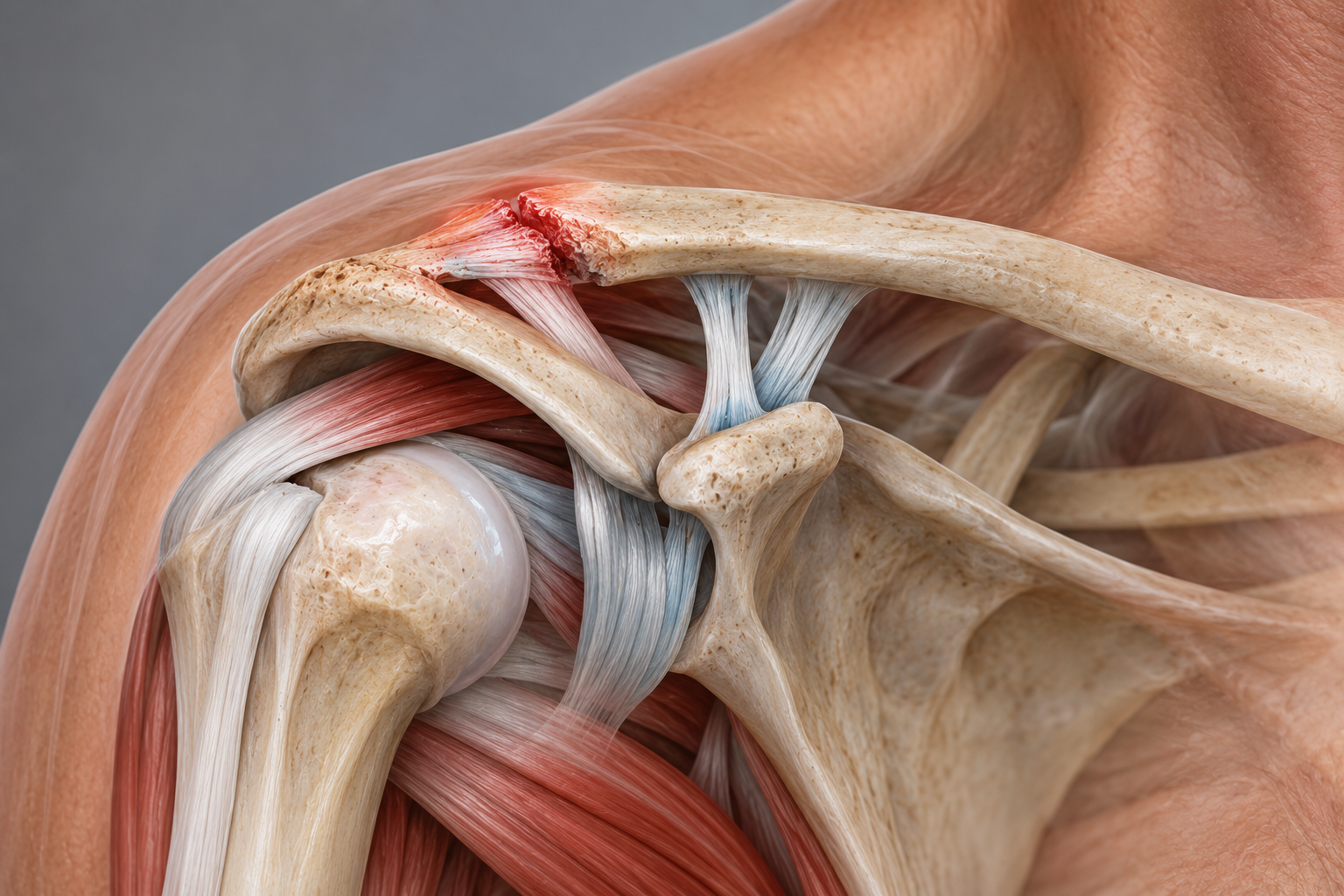
Clinically Relevant Anatomy: The Acromioclavicular Joint
The acromioclavicular joint (AC joint) is a diarthrodial joint that connects the clavicle to the acromion through a fibrocartilaginous, meniscus-like structure. The joint contains an intra-articular synovium and articular cartilage surfaces that permit gliding and rotation. It is characterized by varying angles of inclination in the sagittal and coronal planes, as well as the presence of a disc. Two types of discs have been observed:
Complete disc (very rare).
Meniscoid-like disc (most common).³⁴⁵
Joint Capsule and Ligament Reinforcement
The AC joint is surrounded by a capsule reinforced by:
Superior and inferior capsular ligaments.
Coracoclavicular (CC) ligaments:
Trapezoid ligament attaches anteriorly and laterally on the clavicle.
Conoid ligament attaches posteriorly and medially on the clavicle.
These structures are essential for joint stability:
AC ligament:
Controls horizontal stability in the anteroposterior plane.
CC ligaments:
Maintain vertical stability
Dynamic Stabilization
In addition to the static stabilizers, dynamic muscles contribute, including:
Trapezius: Helps with stability and force transfer across the AC joint.
Deltoid: Supports dynamic control of the joint, particularly during movements involving arm elevation and loading.⁷
This combination of static and dynamic stabilizers allows the AC joint to withstand substantial forces during daily activities and sports. Injury or dysfunction in these structures can impair joint stability and function and lead to painful movement limitations.
Epidemiology and Etiology of Acromioclavicular Joint Injuries
Acromioclavicular joint (AC joint) injuries account for approximately 10% of acute injuries to the shoulder complex. AC joint separations are particularly common in athletes and account for 40% of shoulder injuries in this group. These injuries frequently occur in contact sports and activities with a high risk of falls or direct trauma.⁶
Common Mechanisms of Injury
AC joint injuries usually occur through:
Direct trauma:
Direct force to the lateral point of the shoulder, which may occur from:
A fall directly onto the shoulder.
Impact from another player or object.
This is a common mechanism in sports such as hockey and rugby.⁶⁹
Indirect trauma:
Indirect forces can also cause injury to the AC joint:
For example, a fall onto the elbow may drive the humerus proximally and disrupt the AC joint.
In such cases, the forces affect only the AC ligaments, without involvement of the coracoclavicular ligaments.⁸
Activities and Risk Groups
AC joint injuries are common among athletes participating in activities such as:
Hockey and rugby: Direct contact and falls.
Alpine skiing and snowboarding: Hard falls onto the shoulder or arm.
Football, cycling, and motorsports: High-energy trauma from collisions or crashes.⁹¹⁰
Clinical Significance
Understanding the mechanisms behind AC joint injuries is essential for diagnosis and appropriate treatment planning. Early intervention and appropriate rehabilitation are important for restoring shoulder stability and function, especially in athletes and people participating in high-intensity activities.
Characteristics and Clinical Presentation
Acromioclavicular joint (AC joint) injuries often produce characteristic symptoms that can assist diagnosis and treatment planning. These injuries may cause both local and referred symptoms:
Clinical Symptoms
Pain:
Usually localized to the AC joint, but it may radiate toward the neck and deltoid muscle.
Swelling:
The AC joint may become swollen and tender on palpation.
Positioning of the Upper Extremity:
The arm is often held in adduction to reduce pain.
Depression of the acromion may create the appearance of an elevated clavicle, producing a characteristic deformity.
Classification of AC Joint Injuries
Allman et al. developed a 3-stage classification of AC joint injuries, which was later expanded to a 6-stage model by Rockwood and Green. This classification, known as the Rockwood grading system, is an important tool for:
Identifying the extent of injury.
Selecting appropriate treatment strategies.
Avoiding complications by ensuring that the injury pattern is recognized.
The Rockwood Grading System
Grade 1: Sprain of the AC ligament without visible separation.
Grade 2: Partial rupture of the AC ligament with minor separation.
Grade 3: Complete rupture of the AC and CC ligaments with marked separation.
Grade 4: The clavicle is displaced posteriorly into the trapezius.
Grade 5: Greater displacement with substantial deformity and soft-tissue injury.
Grade 6: The clavicle is displaced inferiorly, beneath the coracoid process.
Correct identification of symptoms and injury grade is essential for selecting the appropriate treatment strategy, whether conservative treatment for lower grades or surgical intervention for more severe injuries. The Rockwood grading system provides a structured approach to assessment and management and helps minimize the risk of long-term complications.
Rockwood Grades for Acromioclavicular Joint Injuries
The Rockwood grading system classifies AC joint injuries according to the extent of injury to the acromioclavicular (AC) and coracoclavicular (CC) ligaments, as well as associated anatomical changes. This system helps determine injury severity and tailor treatment. The following section summarizes the grades, descriptions, and observations/tests:
Grade I:
Description:
Sprain of the AC ligament; both the AC and CC ligaments remain intact.
Observation/Testing:
No clavicular instability is detected during stress testing.
Grade II:
Description:
The AC ligaments are torn, but the CC ligaments remain intact. Often described as a subluxation.
Observation/Testing:
The clavicle is unstable during direct stress testing.
Grade III:
Description:
Complete rupture of both the AC and CC ligaments without major injury to the deltotrapezial fascia. Described as a dislocation.
Observation/Testing:
Deformity with the clavicle appearing elevated due to depression of the acromion. The clavicle is unstable in both the vertical and horizontal directions.
Grade IV:
Description:
The distal clavicle is displaced posteriorly into the trapezius muscle.
Observation/Testing:
Obvious posterior deformity.
Grade V:
Description:
A more severe form of grade III. Complete rupture of both the AC and CC ligaments with injury to the deltotrapezial fascia.
Observation/Testing:
Pseudo-lateral elevation of the clavicle with downward displacement of the scapula.
Grade VI:
Description:
Inferior displacement of the distal clavicle, either subacromially or subcoracoid.
Observation/Testing:
Severe trauma, often accompanied by other major injuries.
Diagnostic Recommendations
Digital measurements are recommended rather than visual assessment alone because they provide greater intra- and interobserver reliability. This improves the accuracy of injury classification and contributes to more precise treatment decisions.¹³
Differential Diagnoses for Acromioclavicular Joint Injuries
Pain and dysfunction in the acromioclavicular joint (AC joint) may often be confused with other shoulder-related conditions. A thorough clinical assessment and appropriate diagnostic approach are required to identify the exact cause. Common differential diagnoses include:
1. Glenohumeral Joint Dislocation:
Description:
Most shoulder dislocations (90%) are anterior and may be associated with concurrent pathologies such as:
Hill-Sachs lesion (compression fracture of the humeral head).
Brachial plexus injury.¹⁴
2. AC Joint Pain From Other Causes:
Osteoarthritis or disc disorder: Degenerative changes may cause pain localized to the AC joint.¹⁵
Distal clavicle osteolysis: A rare but possible cause of pain and dysfunction.¹⁶
AC joint instability: May occur after trauma or degeneration and cause symptoms such as pain and reduced function.¹⁶
3. Rotator Cuff-Related Conditions:
Impingement syndrome: Mechanical compression of the rotator cuff tendons beneath the subacromial arch.¹⁶
Rotator cuff tear: Partial or complete tendon tears can cause substantial pain and functional limitations.¹⁶
4. Adhesive Capsulitis ("Frozen Shoulder"):
Restricted movement in all directions, often accompanied by substantial pain.¹⁶
5. Thoracic Outlet Syndrome:
Compression of the brachial plexus or adjacent vessels at the thoracic outlet may cause radiating pain and neurological symptoms.¹⁶
6. Superior Labral Tears (SLAP Lesions):
Injury to the superior part of the glenoid labrum can cause pain during specific movements, particularly overhead activity.¹⁶
7. Complex Regional Pain Syndrome (CRPS):
A persistent pain condition that may develop following injury or surgery in the shoulder region.¹⁶
8. Other Shoulder-Related Conditions:
Shoulder dislocation: Acute or recurrent dislocation of the glenohumeral joint.⁶
Anterior subluxation of the humerus: Partial displacement of the humeral head relative to the glenoid fossa.⁶
Differential diagnoses for AC joint pain cover a broad range of conditions, from traumatic injuries to degenerative and neurological disorders. Accurate assessment through clinical examination, imaging, and the patient's medical history is necessary to establish the correct diagnosis and ensure appropriate treatment.
Diagnostic Procedures for Acromioclavicular Dislocation
Diagnosis of acromioclavicular (AC) joint dislocation involves both clinical assessment and imaging to confirm the extent of injury. The following procedures are commonly used:
1. Radiography
Primary method: Radiography is the most common diagnostic technique for AC joint injuries.
Limitations:
In grade I injuries radiographs may appear normal because both the AC and CC ligaments appear intact.
Diagnosis of grade I injuries is therefore often based on:
The mechanism of injury.
Clinical tenderness over the AC joint.¹⁷
2. Clinical Tests
Resisted AC Joint Extension Test:
Purpose: To evaluate pain and dysfunction in the AC joint.
Procedure:
The patient sits with the elbow flexed to 90 degrees and the shoulder in 90 degrees of flexion.
The therapist applies resistance while the patient attempts to extend the arm backward.
Positive test:
Pain localized to the AC joint indicates possible injury or inflammation in the area.
When an AC joint injury is suspected, particularly when radiographic findings are insufficient (as in grade I injuries), it is essential to assess the mechanism of injury and perform targeted clinical tests. The combination of radiography and clinical examination provides a comprehensive picture of injury severity and supports appropriate treatment.
Outcome Measures for Acromioclavicular Joint Injuries
Outcome measures are essential for assessing the impact of acromioclavicular joint disorders on a patient’s function, pain, and overall quality of life. These tools are reliable, validated, and commonly used in clinical and research settings to evaluate progress and guide treatment.
1. DASH (Disabilities of the Arm, Shoulder, and Hand) Questionnaire
Purpose:
To evaluate disability of the arm, shoulder, and hand.
Description:
A questionnaire that measures the patient's self-reported functional limitations in daily activities.
Advantages:
Broadly applicable to a range of shoulder and upper-extremity conditions.¹²
2. Simple Shoulder Test (SST)
Purpose:
Assessment of functional disability of the shoulder.
Description:
Consists of 12 questions:
2 related to pain.
7 related to function/strength.
3 related to range of motion.
The questionnaire focuses on the patient's ability to perform specific tasks.¹²
3. Shoulder Pain and Disability Index (SPADI)
Purpose:
Measures the patient's perceived level of pain and disability.
Description:
Consists of two subscales:
Pain: Measures the intensity of shoulder pain.
Disability: Assesses difficulty with daily activities.
Scoring:
Combined score from 0 (no pain or functional difficulty) to 100 (maximum pain and functional difficulty).
Use:
Reliable, valid, and responsive for shoulder pain from musculoskeletal, neurogenic, or unknown causes.¹⁸
4. American Shoulder and Elbow Surgeons (ASES) Questionnaire
Purpose:
Measures functional limitations and pain related to musculoskeletal shoulder conditions.
Description:
Consists of 10 function-related questions rated on a 4-point scale.
Provides a functional score that is particularly useful for clinical follow-up of shoulder pathologies.¹⁸
These outcome measures are important tools for assessing both subjective and objective aspects of shoulder dysfunction. They help to:
Map the patient's perceived symptoms.
Evaluate treatment effectiveness over time.
Support evidence-based practice in the management of acromioclavicular joint injuries.
Examination of the Acromioclavicular Joint (AC Joint)
A thorough examination of the AC joint includes both clinical tests and assessment of the mechanism of injury to confirm the diagnosis and differentiate between injury grades.
1. Clinical Tests
Palpation of the AC joint:
Purpose: Identify tenderness and swelling in the area.
Tenderness over the AC joint is often an early sign of injury.
O’Brien Test:
Purpose: Tensions the posterior capsule and shifts the humeral head posteriorly, thereby stressing the labrum.
Positive test: Pain and weakness suggest a labral injury or AC joint pathology.
Paxinos Sign:
Purpose: Provocation test for AC joint injury.
Procedure:
The therapist applies pressure to the acromion and the lateral end of the clavicle using the thumb and index finger.
Positive test: Pain localized to the AC joint.
Reliability: Walton et al. showed that the Paxinos sign is a useful diagnostic tool. Combined with bone scintigraphy, it provides high diagnostic accuracy.¹¹¹⁹
Stenvers Test 4: Clavicular Roll:
Examines movement of the clavicle relative to the scapula.
Resisted AC Joint Extension Test:
Purpose: Tests pain and instability in the AC joint during resisted extension of the arm.
2. Differentiation Between Injury Types
History and mechanism of injury:
The mechanism of injury (direct trauma or a fall) provides important information about the extent of injury.
Type I injury:
Symptoms: Swelling and pain during arm abduction without visible deformity.
Type II injury:
Symptoms: Tenderness and minor deformity at the AC joint, with pain during all arm movements.
Type III injury:
Symptoms: Clear step deformity at the AC joint, with the patient often supporting the arm close to the body.
3. Diagnostic Significance
The combination of:
Palpation for tenderness.
Provocation tests such as the Paxinos sign.
History and observation of deformity.
These methods provide an accurate assessment of injury grade and differentiate between type I, II, and III injuries.
Clinical Significance
A structured approach to examining the AC joint supports accurate diagnosis and appropriate treatment selection. Early identification of injury severity is essential to prevent long-term complications and optimize rehabilitation.
Medical Treatment of Acromioclavicular Joint Injury
Treatment of AC joint injuries varies according to injury grade and the patient's activity level. A combination of conservative and surgical methods is used depending on injury severity.
Conservative Treatment
Type I and II injuries:
Recommended treatment: Nonoperative approach.
Measures include:
Use of a sling to unload the joint and reduce pain.
Pain relief with ice and anti-inflammatory medication.
Gradual rehabilitation to restore function and strength.
Type III injuries:
Treatment is debated:
Nonoperative treatment may be sufficient in many cases, but there is a high risk of early degenerative changes in the joint.
Surgical intervention may be considered in younger, active patients or when conservative treatment does not provide satisfactory results.
Surgical Treatment
Surgery is often required for more severe injuries:
Type IV and V injuries:
Surgical repair is strongly recommended because of substantial instability and deformity.
Surgical Methods
The four most common surgical approaches are:
AC joint fixation with hook plates:
A plate is implanted to temporarily stabilize the AC joint during healing.
Coracoacromial ligament transfer:
The coracoacromial ligament is transferred to replace or reinforce damaged ligament structures.
Coracoclavicular interval fixation:
Uses implants or other methods to stabilize the interval between the coracoid process and clavicle.
Reconstruction of the coracoclavicular ligaments:
A reconstruction that restores ligament structure and function using autograft, allograft, or synthetic materials.
Choice of treatment depends on several factors:
The patient's age, activity level, and functional demands.
Injury grade according to the Rockwood classification.
The likelihood of satisfactory results with conservative treatment.
For type I and II injuries, conservative treatment is often effective, whereas surgery is recommended for type IV and V injuries and selected type III injuries. Careful assessment helps ensure the optimal approach to restore shoulder stability and function.
Physiotherapy Treatment of Acromioclavicular Joint Injuries
Management of AC joint injuries focuses on reducing pain, restoring function, and preventing complications. Conservative treatment of type I and II injuries follows a structured approach that promotes healing and functional rehabilitation.
Conservative Treatment: Type I and II Injuries
Acute Phase (First 48 Hours)
The POLICE Protocol:
Protection: Protect the joint with a sling to immobilize the shoulder.
Optimal Loading: Gradually introduce load as pain allows.
Ice: Apply ice to reduce swelling and pain.
Compression: Use a bandage or taping for support.
Elevation: Keep the shoulder elevated during rest.
Sling:
Used for immobilization until pain decreases.
Important for providing adequate unloading and reducing stress on the joint.
Taping:
May provide additional support and reduce movement-related pain.
Further Rehabilitation
Timeline for Return to Normal Activities:
Type I injuries: 2–4 weeks.
Type II injuries: 4–6 weeks.
Type III injuries: 6–12 weeks.¹²
Interventions When Progress Is Limited:
If symptoms do not improve within these time frames, intra-articular steroid injections may be considered to relieve inflammation and pain.²³
Rehabilitation Protocols
Although evidence for standardized rehabilitation protocols is limited, Reid et al. developed a best-practice guideline following a systematic review of existing practice.¹²
Clinical Significance
A conservative approach to AC joint injuries, particularly type I and II injuries, is often effective when the POLICE protocol and gradual rehabilitation are followed. For patients with slow recovery or persistent symptoms, more advanced measures such as steroid injections may be considered. Structured follow-up supports optimal functional recovery and helps prevent complications.
Physiotherapy Approach: Acute and Recovery Phases for AC Joint Injuries
Effective rehabilitation of the acromioclavicular joint (AC joint) follows a structured approach tailored to injury grade and the patient's functional level. Rehabilitation is divided into two main phases: the acute phase and the recovery phase.
Acute Phase
Goals:
Reduce pain and inflammation.
Maintain and gradually improve range of motion (ROM).
Preserve muscle activation without aggravating the injury.
Range-of-Motion (ROM) Exercises:
Glenohumeral joint (GHJ
Internal/external rotation and flexion as tolerated:
Towel pulls.
Pendulum exercises.
Supported range-of-motion exercises using an L-bar:
GH joint at 30°–45° abduction and 30°–40° forward flexion.
Scapular movements:
Protraction, retraction, elevation, and depression.
Soft-Tissue Mobilization:
Focus on reducing tightness in the pectoralis minor with stretching.
Sleeper stretch for the posterior GH joint.
Isometric exercises:
Performed at multiple angles with submaximal, pain-free intensity.
Closed Kinetic Chain Exercises (Unloaded):
Hand support in different planes and at different levels to control scapular position.
Examples:
Wall push.
Scapular clock.
Elbow-supported rotational exercises.
Recovery Phase
Goals:
Restore full range of motion.
Strengthen stabilizers and upper-extremity musculature.
Integrate functional and sport-related movements.
Range of Motion (ROM):
Focus on full flexion, horizontal adduction, and internal/external rotation at 90° abduction.
Capsular stretching (e.g. sleeper stretch).
Closed kinetic chain exercises:
Gradual increase in loading:
Hand support with active arm elevation and rotation.
Progress to activities that include unstable surfaces, such as a ball or planks.
Open kinetic chain exercises (OKC):
Combined with closed-chain exercises:
Wall push with trunk and lower-extremity involvement.
Pushing in the scapular plane.
Kinetic Chain Training:
Scapular retraction: Trunk and hip extension, e.g. low-row exercises.
Scapular protraction: Trunk and hip flexion, e.g. punches.
Trunk rotations: Combined pulling and rotational exercises, e.g. upper cuts.
Deltotrapezial complex:
Exercises using cables, shrugs, and abduction at varied angles.
Plyometric Exercises and Sport-Related Movements:
Medicine-ball throws and catches:
Dynamic stretch-shortening cycle.
Sport-related activities:
Example: Side-to-side overhead throwing for athletes who use the upper extremities.
Return to Sport
Grade I: 2–4 weeks.
Grade II: 4–8 weeks.
Grade III: 6–12 weeks, depending on progression and stability.
This gradual approach balances protection of the joint with promotion of functional recovery and is individualized to achieve optimal outcomes.
Postoperative Rehabilitation for Type V and VI AC Joint Injuries
Type V and VI injuries of the acromioclavicular joint (AC joint) are considered severe and usually require surgical repair. Following surgery, physiotherapy plays an important role in restoring shoulder function, reducing pain, and preventing complications.
Goals After Surgery
Control pain and swelling.
Protect the AC joint repair.
Promote wound healing.
Begin early shoulder movement.
Postoperative Management
Pain Relief and Reduction of Swelling:
Use cold compresses for 20 minutes several times per day to reduce pain and swelling.
Pendulum Exercises:
Remove the sling several times daily to perform pendulum movements:
Lean forward and allow the arm to swing passively.
Perform for 3–5 minutes every 1–2 hours.
Active Movement of Distal Joints:
Remove the sling several times daily to move the elbow, wrist, and hand.
Hygiene:
To wash under the operated arm, bend forward at the waist so the arm hangs passively away from the body. This position is safe and provides space for hygiene.
Variable Protocols for Movement and
sling use
Sling Use:
Some surgeons recommend continuous sling use for up to 6 weeks to protect the joint.
Others allow sling use as needed, with active movement up to 90 degrees during the first two weeks.
Progression of Movement:
Early movement is often limited to pain-free ROM.
Gradual increase in range of motion and strength over time.
Rehabilitation Protocols
Postoperative rehabilitation follows similar principles to those used for type I and II injuries:
ROM exercises:
Focus on gradual restoration of range of motion.
Strength Training:
Progress from isometric exercises to dynamic strengthening exercises.
Long-Term Follow-Up:
Rehabilitation should continue until full strength and mobility are restored to prevent persistent pain and instability.
Surgical Treatment Versus Conservative Treatment
For type III injuries:
Studies, including Bannister et al., have shown that conservative treatment often provides faster restoration of shoulder kinematics with fewer complications than surgery.²⁵²⁶
For type IV and V injuries:
Although surgery is preferred, successful manual reductions converting type IV injuries to type II have been reported.²⁷
Clinical Significance
Postoperative management of AC joint injuries requires an individualized approach based on injury grade, surgical protocol, and patient needs. By combining pain control, gradual progression of movement and strength, and long-term follow-up, patients can achieve optimal function and reduce the risk of complications such as pain and instability.
Sources
Codsi JM. The painful shoulder: when to inject and when to refer. Cleveland clinic journal of medicine 2007; 74(7): 473-482. (level of evidence 4)
Heijmans E, Eekhof J; Neven AK. Acromioclaviculaire luxatie, huisarts & wetenschap, november 2010
Saccomanno MF. Acromioclavicular joint instability: anatomy, biomechanics and evaluation. Joints 2014; 2(2): 87–92.
De Palma AF. Surgical anatomy of the acromioclavicular and sternoclavicular joints. Surg Clin North Am. 1963;43:1541–1550.
Salter EG, Jr, Nasca RJ, Shelley BS. Anatomical observations on the acromioclavicular joint in supporting ligaments. Am J Sports Med 1987;15(3):199-206.
Magee DJ, Zachazewski JE, Quillen WS. Pathology and Intervention in Musculoskeletal Rehabilitation.fckLRElsevier Health Sciences, 2008.
Suezie K, Blank A, Strauss E. Management of Type 3 Acromioclavicular Joint Dislocations Current Controversies. Bulletin of the Hospital for Joint Diseases 2014; 72(1): 5360.
Beim GM. Acromioclavicular joint injuries. Journal of Athletic Training 2000;35(3):261-267.
Johansen JA, Grutter PW, McFarland EG, Petersen SA. Acromioclavicular joint injuries: indications for treatment and treatment options. J Shoulder Elbow Surg 2011;20:70-82.
Culp LB, Romani WA. Physical Therapist Examination, Evaluation, and Intervention Following the Surgical Reconstruction of a Grade III Acromioclavicular Joint Separation. Journal of the American physical therapy association 2006; 86:857-869.
Micheli LJ. Encyclopedia of Sports Medicine. London: SAGE Publications, 2010.
Reid D, Polson K, Johnson L, Acromioclavicular Joint Separations Grades I–III A Review of the Literature and Development of Best Practice Guidelines. Sports Med. 2012; 42(8): 681-696.
Schneider MM, Balke M, Koenen P, Fröhlich M, Wafaisade A, Bouillon B, Banerjee M. Inter- and intraobserver reliability of the Rockwood classification in acute acromioclavicular joint dislocations. Knee Surg Sports Traumatol Arthrosc. 2016; 24(7): 2192-6.
Nepola VJ, Newhouse EK, Recurrent shoulder dislocation. The iowa orthopaedic journal 1993; 13: 97-106
Robb AJ, Howitt S, Conservative management of a type III acromioclavicular separation: a case report and 10-year follow-up. Journal of Chiropractic Medicine 2011; 10: 261–271.
Fraser-Moodie JA, Shortt NL, Robinson CM. Injuries to the acromioclavicular joint. J Bone Joint Surg. 2008 ;90-B: 697-707.
4. Johansen JA, Grutter PW, McFarland EG, Petersen SA. Acromioclavicular joint injuries: indications for treatment and treatment options. J Shoulder Elbow Surg. 2011;20 p.S70-82
Harris KD, Deyle GD, Gill NW, Howes RR. Manual Physical Therapy for Injection-Confirmed Nonacute Acromioclavicular Joint Pain. Journal of orthopaedic & sports physical therapy 2012; 42(2): 66-80.
Walton J, Mahajan S, Paxinos A, Marshall J, Bryant C, Shnier R, Quinn R, Murell GAC. Diagnostic Values of Tests for Acromioclavicular Joint Pain. The Journal Of Bone & Joint Surgery 2004; 86-A (4): 807-812.
Culp LB, Romani W. Physical Therapist Examination, Evaluation, and Intervention Following the Surgical Reconstruction of a Grade III Acromioclavicular Joint Separation. Journal of the American physical therapy association 2006; 86:857-869.
Nabil Ebraheim. Examination Of The AC Joint - Everything You Need To Know - Dr. Nabil Ebraheim. Available from https://www.youtube.com/watch?v=daPnkXo03yM&t=1s
Johansen JA, Grutter PW, McFarland EG, Petersen SA. Acromioclavicular joint injuries: indications for treatment and treatment options. J Shoulder Elbow Surg. 2011;20: S70-82
Hootman JM. Acromioclavicular Dislocation: Conservative or Surgical Therapy. Athl Train. 2004; 39(1):10–11.
Physio Fitness | Physio REHAB | Tim Keeley "Weight-lifter's shoulder" pain from an unstable AC joint | Feat. Tim Keeley | No. 27 | Physio REHAB. Available from https://www.youtube.com/watch?v=aLj--YqCXhw&t=2s
Bannister GC, Wallace WA, Stableforth PG, Hutson MA. The management of acute acromioclavicular dislocation. A randomised prospective controlled trial. Bone Joint Surg Br. 1989; 71(5): 848-850.
Nissen CW, Chatterjee A. Type III acromioclavicular separation: results of a recent survey on its management. Am J Orthop (Belle Mead NJ). 2007 Feb. 36(2):89-93.
Johansen JA, Grutter PW, McFarland EG, Petersen SA. Acromioclavicular joint injuries: indications for treatment and treatment options. J Shoulder Elbow Surg. 2011;20 p.S70-82
Johansen JA, Grutter PW, McFarland EG, Petersen SA. Acromioclavicular joint injuries: indications for treatment and treatment options. J Shoulder Elbow Surg. 2011; 20: S70-82
Glick JM, Milburn LJ, Haggerty JF, Nishimoto D. Dislocated acromioclavicular joint: follow-up study of 35 unreduced acromioclavicular dislocations. Am J Sports Med 1977; 5: 264-70.


