

Acute Myeloid Leukemia (AML) – Blood Cancer

Acute myeloid leukemia (AML) is the most common form of acute blood cancer in adults and accounts for approximately 80% of acute leukemias in this group. AML is a malignant blood disorder that affects the bone marrow, where normal blood cells are gradually replaced by immature and dysfunctional cells called myeloblasts. These cells arise from myeloid stem cells, which normally develop into red blood cells, white blood cells and platelets. In AML, this process is disrupted, and the myeloblasts remain immature and unable to perform the functions of healthy blood cells. The bone marrow consequently becomes filled with leukemic cells, impairing the production of healthy erythrocytes, leukocytes and thrombocytes.
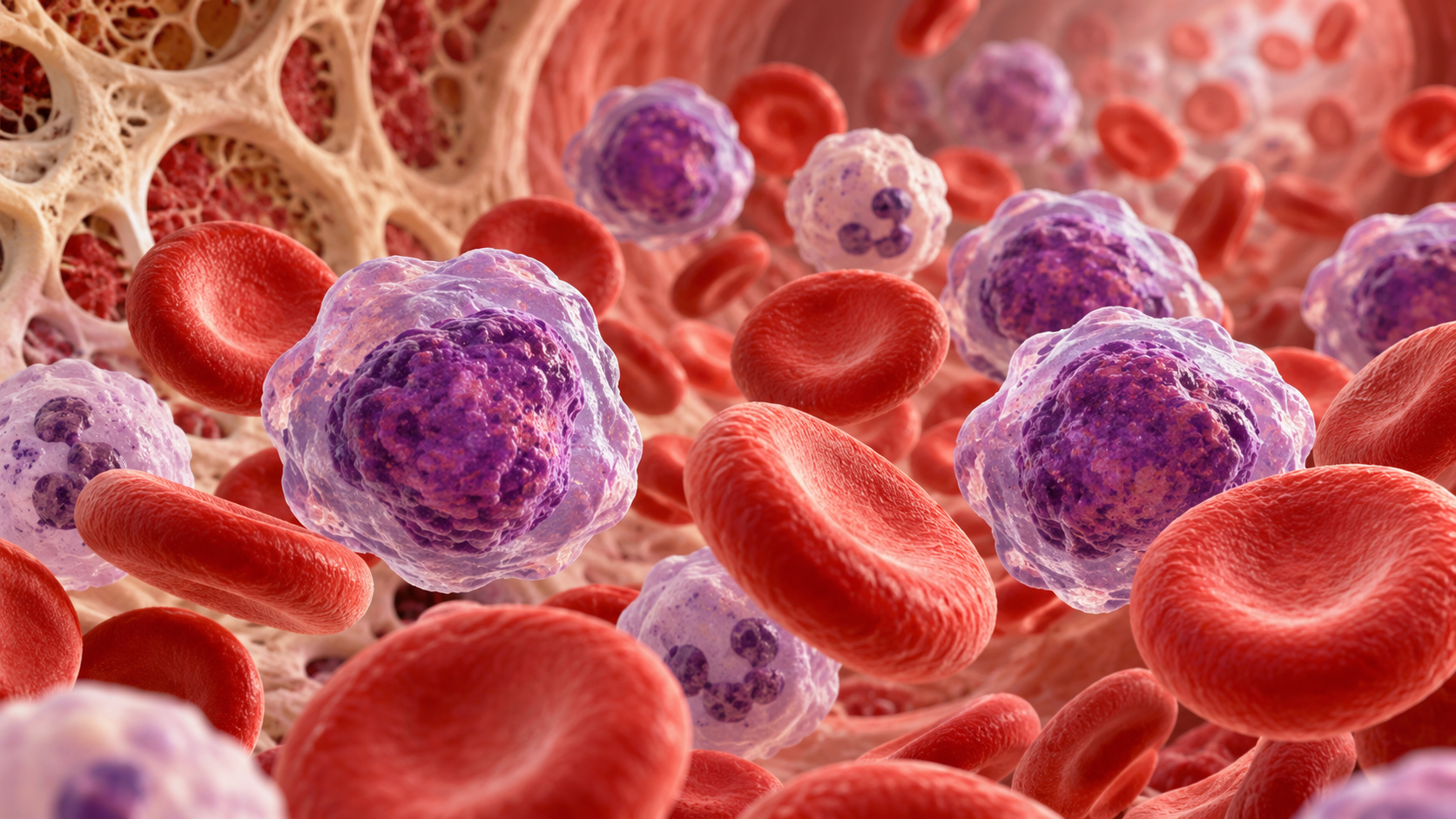
AML causes progressive failure of hematopoiesis, which in turn leads to serious symptoms related to anemia, infections and bleeding tendency. Despite major therapeutic advances, prognosis remains poor, particularly among older adults. In patients over 60 years of age, modern treatment regimens have produced survival rates of up to 15%, while patients under 60 have an estimated cure rate of around 40%¹.
Epidemiology
AML has an incidence of approximately 4.2 new cases per 100,000 people per year. In the United States, more than 20,000 new cases are diagnosed annually, and the disease most commonly affects people with a mean age of approximately 65 years at diagnosis¹. AML is more common in men than women, with a sex distribution of approximately 5:3¹. It is also more prevalent among non-Hispanic White populations, and risk rises sharply with age: people over 70 years of age are 12 times more likely to develop AML than younger adults³. Although most cases occur in adults, children account for approximately 20% of the AML population.
Clinical presentation
Symptoms of AML primarily arise from impaired production of normal blood cells and vary depending on which cell lines are most affected. Reduced neutrophil production increases susceptibility to infection, while thrombocytopenia predisposes patients to spontaneous or disproportionate bleeding. Many patients report bleeding after minor trauma, bleeding gums, unusually heavy menstrual bleeding or intermenstrual bleeding. Petechiae, small purple skin hemorrhages, are a common clinical finding.
Anemia caused by reduced erythropoiesis produces symptoms such as fatigue, dyspnea on mild exertion and general loss of energy. Patients may also develop leukemia cutis, characterized by multiple purple papules or a more diffuse rash. The spleen may become enlarged (splenomegaly) in approximately half of patients. Bone pain may also occur, particularly in the ribs, sternum and tibia, because of bone-marrow infiltration.
In older patients, AML may cause more nonspecific symptoms such as confusion, persistent weakness and pallor, and the disease is often diagnosed later in its course than in younger patients.
Comorbidities
AML is often accompanied by several hematological and systemic conditions that may worsen prognosis and complicate treatment:
Thrombocytopenia: A low platelet count increases the risk of spontaneous bleeding and impairs hemostasis.
Neutropenia: A reduced neutrophil count weakens immune defense and increases the risk of life-threatening infections.
Diabetes mellitus: A metabolic disorder characterized by insulin resistance and/or reduced insulin production, which may increase susceptibility to infection and impair wound healing.
Systemic lupus erythematosus (SLE): An autoimmune connective-tissue disease that attacks healthy tissue and may increase the risk of secondary malignancies.
Multiple sclerosis (MS): An inflammatory disease of the central nervous system in which the immune system attacks myelin around nerve cells.
Rheumatoid arthritis (RA): A chronic autoimmune disease affecting the joints that shares several immunological features with AML⁴.
Diagnostic tests and laboratory values
AML is diagnosed through a combination of clinical assessment, laboratory testing and bone-marrow examination. Diagnosis is based on the presence of myeloblasts in the blood and/or bone marrow, and the disease is characterized by extensive impairment of hematopoiesis.
History and clinical examination: Focuses on symptoms such as fatigue, bleeding, infections and weight loss.
Blood tests: Typical findings include an elevated leukocyte count with immature cells (myeloblasts), together with anemia and thrombocytopenia.
Bone-marrow aspiration and biopsy: The diagnostic gold standard, allowing morphological and cytogenetic features of leukemic cells to be analyzed.
Immunophenotyping: Used to classify the AML subtype by identifying characteristic surface antigens on leukemic cells²,³.
Causes and risk factors
Although the exact etiology of AML is not fully understood, several risk factors are known:
Smoking: A known risk factor for several cancers, including hematological malignancies.
Benzene exposure: Long-term exposure to this industrial chemical significantly increases risk.
Previous cancer treatment: In particular, treatment with alkylating cytotoxic agents and platinum compounds may cause secondary AML.
Radiation: Previous radiation exposure, especially at high doses, is associated with increased risk.
Pre-existing hematological disorders: Blood disorders such as polycythemia vera, essential thrombocythemia and idiopathic myelofibrosis increase AML risk.
Genetic syndromes: Fanconi anemia, Down syndrome, Bloom syndrome, Li-Fraumeni syndrome, Kostmann syndrome and trisomy 8 are among the genetic conditions associated with AML.
Family history: Having a close relative with AML may indicate increased genetic susceptibility.
Sex: Men have a higher incidence of AML than women⁵.
Systemic involvement
AML can affect several organ systems as a result of leukemic-cell infiltration, bone-marrow failure and increased metabolic activity:
Central nervous system: Infiltration may lead to headache, disorientation and, in some cases, neurological deficits.
Anemia and hypoxia: May cause pallor, dizziness, fatigue and shortness of breath.
Infections and sores in the mouth/throat: These occur because immature leukocytes do not effectively fight pathogens, resulting in impaired immunity.
Increased metabolic activity: The high proliferation rate of leukemic cells increases energy expenditure and may cause weight loss, weakness and catabolism.
Hyperuricemia and renal failure: Breakdown of large numbers of leukocytes can cause uric-acid accumulation, which may damage the kidneys and lead to uremia.
Hepatosplenomegaly: Leukemic cells may accumulate in the spleen and liver, causing enlargement of these organs.
Lymphadenopathy and skeletal pain: Infiltration of lymph nodes and bone marrow may cause swelling and pain³.
Medical treatment of acute myeloid leukemia
Treatment of acute myeloid leukemia (AML) is complex and often intensive, and chemotherapy is the primary therapeutic approach. The goals are to eliminate leukemic cells, restore normal hematopoiesis and achieve sustained remission. Treatment is commonly divided into induction, consolidation and, for selected disease subtypes, maintenance phases, depending on disease characteristics and response to treatment.
Induction phase
The goal of this initial phase is to reduce the number of leukemic cells in the blood and bone marrow. Intensive chemotherapy is given to achieve complete remission, defined by normalization of blood counts and absence of detectable leukemia in the bone marrow. Patients are monitored closely for treatment response and complications, particularly infection and bone-marrow failure.
Consolidation phase
After recovery from induction, additional chemotherapy is given in one or more cycles to eliminate residual microscopic leukemic cells that were not detectable during remission. This phase is important for reducing the risk of relapse.
Maintenance phase
This phase is relevant for selected AML subtypes and involves low-dose therapy given over months or years. The aim is to reduce the risk of recurrence by suppressing minimal residual disease over time.
Standard treatment approaches for AML
Chemotherapy
Chemotherapy is the foundation of AML treatment and works by killing cancer cells or inhibiting their ability to divide. Treatment may be systemic, given as tablets, intravenous injection or intramuscular injection, thereby acting throughout the body. When leukemic cells have spread to the central nervous system, intrathecal chemotherapy may be used, with medication delivered directly into the cerebrospinal fluid. In selected situations, regional chemotherapy may also be used, delivering treatment directly to an affected region.
Radiation therapy
Radiation therapy is used less frequently in AML but may be considered for localized disease, such as leukemic masses (myeloid sarcoma). External-beam radiation is directed at the body from outside, while internal radiation therapy involves placing radioactive material near the affected area. The approach is selected according to AML subtype and disease distribution.
Stem-cell transplantation
Stem-cell transplantation may be used after high-dose chemotherapy and involves infusion of the patient's own (autologous) or a donor's (allogeneic) blood stem cells after treatment. Before transplantation, the patient's bone marrow is suppressed or destroyed with intensive chemotherapy, sometimes combined with radiation, after which the infused stem cells establish new hematopoiesis. This may be appropriate for patients at high risk of relapse or those who do not respond adequately to standard chemotherapy.
Targeted therapy
Targeted therapy is a newer and more specific approach that uses drugs or biological agents directed at particular structures or signaling pathways in leukemic cells. Monoclonal antibodies are one example and may bind to antigens on leukemic cells, leading to cell destruction or inhibition of growth. These treatments are often administered intravenously, either alone or in combination with chemotherapy. Several clinical trials are ongoing to optimize targeted therapy in AML²,³.
Treatment is individualized according to factors such as age, general health, cytogenetic profile and response to initial therapy. In older patients and those with comorbidities, treatment often needs to be adjusted to reduce toxicity, with greater emphasis on quality of life and symptom relief.
Physiotherapy in acute myeloid leukemia
Physiotherapy can play an important role in the management of patients with acute myeloid leukemia (AML), both during and after cancer treatment. Goals include maintaining physical function, reducing symptom burden, improving quality of life and counteracting adverse effects associated with cytotoxic therapy and prolonged inactivity. Treatment is adapted to the patient's general condition and hematological status, with particular emphasis on safety and symptom management.

Aerobic exercise
Aerobic physical activity has documented benefits for fatigue, mental health and overall function in people with cancer. Studies suggest that as little as 12 minutes of walking five times per week may reduce cancer-related fatigue, anxiety, depression and symptom burden while helping maintain cardiovascular health⁶. Activity should be adjusted to daily capacity and may also help prevent complications of prolonged bed rest such as pressure injuries, constipation, pulmonary secretion retention and muscle atrophy.
Strength training
A combination of strength and endurance exercise three times per week, with two daily sessions of 30 minutes, has shown positive effects on endurance, overall fatigue, depression and inflammatory biomarkers. Exercises may be simple and functional, including active movement against gravity, grip-strength exercises and exercises using resistance bands or light resistance for the upper and lower limbs⁷. The goal is to maintain muscle strength and function throughout treatment.
Pain management
Pain may be a significant problem because of both the disease itself and its treatment. Modalities such as TENS, heat and cold packs, massage and optimal positioning may be used for symptom relief. The choice of method should be based on patient preference and clinical assessment.
Stretching and joint mobility
Daily stretching may help prevent contractures and joint stiffness in patients with reduced mobility. Active and passive range-of-motion exercises, together with static stretching, may be incorporated, and orthoses or splints may be considered to maintain joint position. Stretching can be performed at least five days per week, with each muscle group stretched four times for 12–15 seconds per session.
Balance training and neurological rehabilitation
A common adverse effect of chemotherapy is chemotherapy-induced peripheral neuropathy (CIPN), which can cause reduced sensation, pain and impaired balance. Balance exercises and neuromuscular training should therefore be integrated early when appropriate to reduce fall risk and maintain safe mobility.
The comprehensive role of cancer rehabilitation
Physiotherapists should be part of the multidisciplinary team and contribute individualized exercise planning, patient education and functional assessment. The patient's preferences, treatment phase and hematological values must always be considered before physical activity is initiated.
Important precautions for AML and physiotherapy
Avoid exercise in the presence of severe fatigue, marked anemia or neurological symptoms such as ataxia.
When the white blood-cell count is low (< 3.5 × 10⁹/L), the patient may need to avoid public environments and group-based exercise because of increased infection risk.
Neuropathy increases fall risk; implement preventive measures and monitor balance.
Be aware of adverse effects from medications, including cytotoxic drugs and corticosteroids.
Follow universal hygiene precautions because patients with AML may be immunocompromised⁸.
Sources:
Vakiti A, Mewawalla P. Cancer, acute myeloid leukemia (AML, erythroid leukemia, myelodysplasia-related leukemia, BCR-ABL chronic leukemia). InStatPearls [Internet] 2019 Nov 28. StatPearls Publishing.:https://www.ncbi.nlm.nih.gov/books/NBK507875/
National Cancer Institute. Adult Acute Myeloid Leukemia Treatment. National Cancer Institute at the National Institute of Health. December 26, 2013. http://www.cancer.gov/cancertopics/pdq/treatment/adultAML/Patient/page1,
Goodman CC, Fuller KS: Pathology: implications for the physical therapist, ed 3, Philadelphia, 2009, WB Saunders.
Goodman C, Snyder T. Differential Diagnosis for Physical Therapists: Screening for Referral. Saunders, St. Louis, Missouri. 5th edition. 2013.
American Cancer Society. What are the risk factors for acute myeloid leukemia? American Cancer Society. February 7, 2014. http://www.cancer.org/cancer/leukemia-acutemyeloidaml/detailedguide/leukemia-acute-myeloid-myelogenous-risk-factors.
Chang PH, Lai YH, Shun SC, et al. Effects of a walking intervention on fatigue-related experiences of hospitalized acute myelogenous leukemia patients undergoing chemotherapy: a randomized controlled trial. J Pain Symptom Manage. 2008;35(5):524-34.
Battaglini CL, Hackney AC, Garcia R, Groff D, Evans E, Shea T. The effects of an exercise program in leukemia patients. Integr Cancer Ther. 2009;8(2):130-8.
Breastcancer.org. Exercise during and after chemotherapy or targeted therapies. Breastcancer.org. January 26, 2014.http://www.breastcancer.org/tips/exercise/treatment/chemo_targeted.


