

Adductor Tendinopathy

Adductor tendinopathy refers to a group of conditions that develop in and around the adductor tendons as a result of chronic overload[1]. The condition is common among athletes and other highly active people whose activities repeatedly load the adductor muscles.
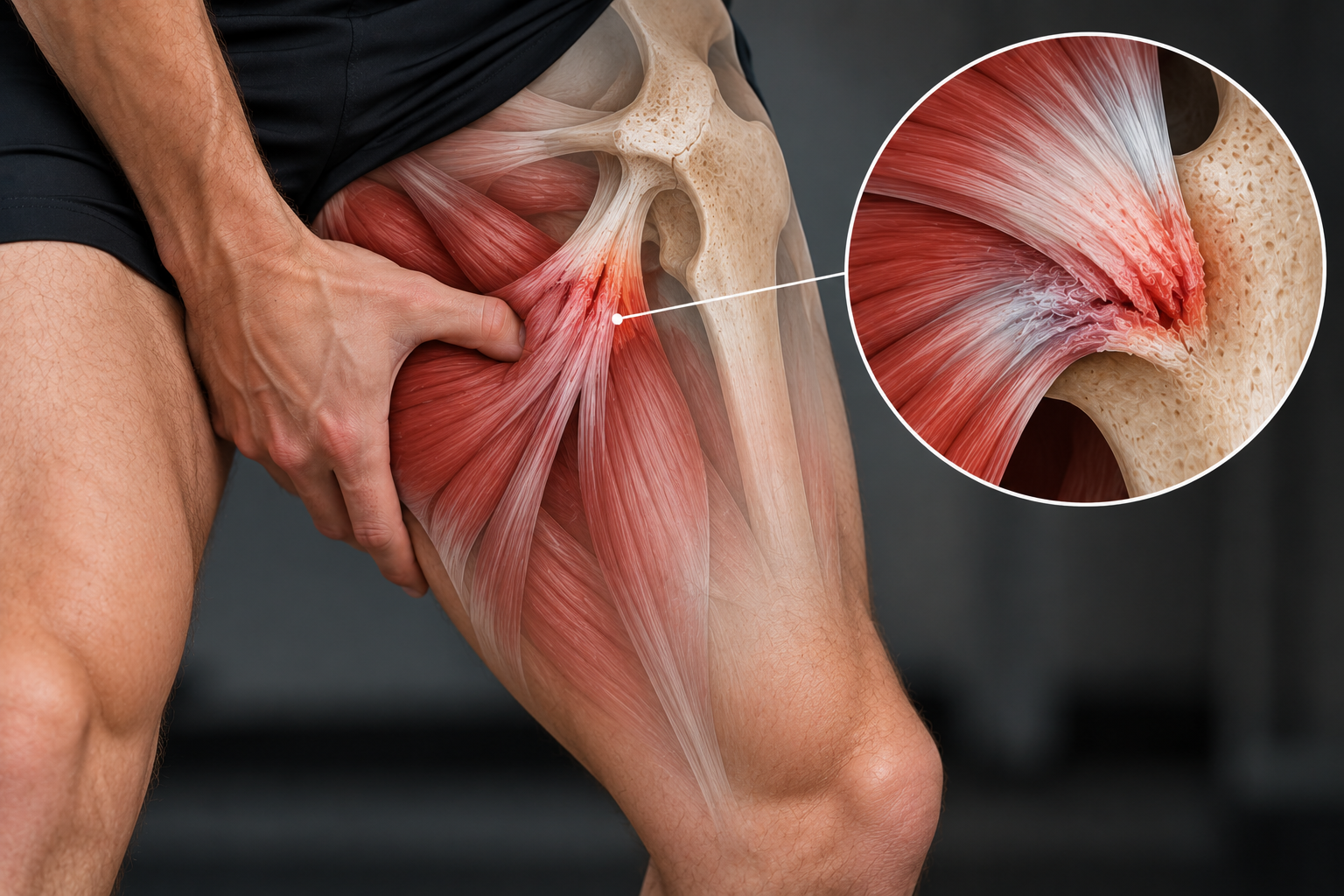
Pathophysiology
Histopathological changes
At the cellular level, the following changes may be observed in the tendon:
Collagen separation and degeneration: This involves structural changes in the collagen fibers that weaken the integrity of the tendon[1].
Macroscopic changes
Tendon thickening: Chronic stress can lead to increased tendon volume as a compensatory response.
Loss of mechanical properties: The tendon's ability to tolerate load is reduced.
Pain: This is a central symptom that can limit functional capacity[2].
Inflammation and terminology
Although inflammation was previously believed to play a central role, more recent research has shown that inflammatory cells are often absent around the lesion. This has led to the term "tendinitis" being replaced by "tendinopathy" to better reflect the largely non-inflammatory nature of the condition[3].
Anatomy and function
The adductor group consists of five muscles that contribute to pelvic stabilization and perform adduction, moving the legs toward the body's midline:
Long adductors
Gracilis
Adductor Magnus
These muscles extend from the pelvis toward the knee.
Short adductors
Pectineus
Adductor Brevis
Adductor Longus
These extend from the pelvis to the femur[4].
The adductor muscles are important for stabilizing the pelvis during walking, running and other activities that require control of the hip joint.
Adductor tendinopathy can cause pain and reduced function in the hip and thigh, affecting daily activities and athletic performance. Understanding the pathophysiology and anatomy is important for diagnosis and treatment.
Clinically relevant anatomy of the adductor muscles
The adductor muscles are located between the flexor and extensor compartments of the thigh. They play an important role in stabilizing the pelvis and in movements requiring adduction, such as crossing the legs. The muscles extend from the lower pelvis to the femur and, in some cases, to the region around the knee.
Overview of the adductor muscles
Adductor Magnus
The largest muscle in the group, positioned posteriorly relative to the other adductors.
Divisions:
Adductor portion:
Origin: Inferior pubic ramus and ischial ramus.
Insertion: Linea aspera of the femur and the medial epicondyle through its tendinous attachment.
Function: Adduction and flexion of the thigh.
Hamstring portion:
Origin: Ischial tuberosity.
Insertion: Adductor tubercle and medial supracondylar line of the femur.
Function: Adduction and extension of the thigh.
Adductor Longus
A large, flat, fan-shaped muscle that forms part of the medial border of the femoral triangle.
Origin: Superior pubic ramus and pubic symphysis.
Insertion: Linea aspera of the femur.
Function: Adduction and medial rotation of the thigh.
Special feature: Distally, it forms an aponeurosis that extends toward the vastus medialis muscle.
Adductor Brevis
Located deep to adductor longus.
Origin: Inferior pubic ramus.
Insertion: Posterior aspect of the linea aspera.
Function: Adduction of the thigh.
Gracilis
The only biarticular muscle in the group.
Origin: Inferior border of the pubic symphysis.
Insertion: Medial surface of the tibia, where it contributes to the pes anserinus together with the sartorius and semitendinosus tendons.
Function: Adduction of the thigh and flexion of the knee.
Special feature: The most superficial muscle of the adductor group.
The adductor muscles are essential for pelvic stabilization during standing and walking. Injury or dysfunction of these muscles, including adductor tendinopathy, can cause pain, reduced mobility and impaired hip strength. An understanding of the anatomy and function of these muscles is important for accurate diagnosis and treatment.
Etiology of adductor tendinopathy
Adductor tendinopathy often develops because of repetitive loading or sudden changes in activity, particularly in athletes. The condition is multifactorial and may be influenced by biomechanical, training-related and individual factors.
Common causes of adductor tendinopathy
Sport-related factors
The adductor muscles are heavily used in sports such as running, football, horse riding, gymnastics and swimming. These activities can place substantial stress on the adductor tendons, particularly when there are:
Repetitive movements: Repeated performance of the same movement, as in running and swimming, can lead to microtrauma in the tendon[4].
Frequent changes of direction: Sports such as football or basketball increase loading of the adductor tendons during rapid movements and cutting maneuvers.
Overstretching: Excessive stretching of the adductor tendons can cause acute injury or contribute to chronic overload[4].
Training- and load-related factors
Sudden increase in training volume: A rapid increase in training intensity or volume may exceed the adductor tendon's capacity to adapt.
Change in type of training: Introduction of new movements or activities that challenge the adductor muscles in unfamiliar ways.
Biomechanical and physiological risk factors: Adductor tendinopathy often develops through a combination of factors that influence tendon loading:
Leg-length discrepancy: Although the threshold for a clinically significant discrepancy is not clearly defined, differences in leg length may alter gait and place additional stress on the adductor muscles.
Poor movement patterns: Suboptimal technique during physical activity can overload the adductor tendons.
Muscle imbalance:
Differences in muscle length or strength around the hip, thigh or abdominal region may adversely affect load distribution.
Weakness in the lower-extremity or abdominal musculature may contribute to the development of tendinopathy.
Other risk factors
Insufficient warm-up: This may increase injury risk because of reduced muscle readiness and flexibility.
Inactivity and fatigue: Reduced muscular capacity may result in overload when activity is resumed.
Age-related weakening: Natural degeneration of tendon tissue with increasing age.
Obesity: Excess body weight may increase loading on the adductor tendons.
Genetics: Inherited factors may influence the strength and elasticity of connective tissue.
Unclear pathology
Although many factors may contribute to the development of adductor tendinopathy, the exact pathology remains incompletely understood. A comprehensive approach that considers biomechanical, training-related and individual factors is therefore important[4].
Characteristics of adductor tendinopathy
Adductor tendinopathy typically presents with pain in the groin region and is often associated with loading or use of the adductor muscles. Symptoms may range from gradually developing discomfort to episodes of acute pain depending on severity.
Typical symptoms
Groin pain
Often experienced as a deep, aching pain during:
Palpation of the adductor tendons.
Active adduction, bringing the leg toward the midline.
Passive movement of the affected leg into adduction[4].
Pain development
Pain may develop gradually as a result of chronic overload.
Acute, sharp pain may occur with overstretching or sudden loading.
Swelling or a lump
Swelling or a palpable lump may be present in the adductor muscles as a result of tendon injury or irritation.
Stiffness in the groin region
Often experienced as reduced flexibility or discomfort when attempting to stretch the adductor muscles.
Functional limitations
In severe cases, the patient may experience:
Difficulty contracting or stretching the adductor muscles.
Restrictions in daily physical activities because the tendon can no longer tolerate repeated loading[4].
Differential diagnoses for adductor tendinopathy
Groin pain can have many different causes, including musculotendinous problems, neurological conditions and pathology involving internal structures in the region. Adductor tendinopathy can be difficult to distinguish from other conditions, particularly osteitis pubis, which often presents with overlapping symptoms. A thorough clinical assessment and appropriate diagnostic evaluation are essential for identifying the underlying cause.
Common differential diagnoses
Osteitis pubis
Chronic inflammation or irritation of the pubic symphysis and surrounding structures.
Characterized by groin pain, often bilateral, with stiffness and tenderness over the pubic symphysis.
Can be difficult to distinguish from adductor tendinopathy without imaging.
Sports hernia or inguinal hernia
Weakness or tearing in the inguinal region that causes pain during activity.
Pain may worsen with coughing, sneezing or straining.
Iliopsoas bursitis
Inflammation of the iliopsoas bursa can cause groin pain, often together with symptoms of snapping hip.
Stress fracture
Stress fractures of the pelvis, particularly the pubic bone or femoral neck, may cause gradually developing groin pain.
Often associated with overload or repetitive activity.
Avulsion fracture
Avulsion of the adductor tendon attachment from the pelvis may cause acute pain, particularly in young athletes.
Nerve compression
Compression of nerves such as the obturator or ilioinguinal nerve can cause pain that may be mistaken for adductor-related pain.
Snapping hip syndrome
Characterized by an audible or palpable snapping sensation around the hip, often associated with pain.
Chronic prostatitis
May cause referred pain to the groin and should be considered in men with concurrent urinary symptoms.
Diagnostic procedures for adductor tendinopathy
Accurate diagnosis of adductor tendinopathy requires careful assessment of symptoms combined with clinical examination and, when appropriate, imaging.
Clinical assessment by a physiotherapist
Physiotherapists can establish an objective clinical diagnosis based on:
History: Assessment of symptoms, pain-provoking activities and previous injuries.
Physical examination:
Palpation of the adductor tendons to identify pain or swelling.
Functional tests that reproduce pain during hip adduction.
Advanced imaging
In more complex cases, or when clinical examination is insufficient, the following diagnostic tools may be used:
Ultrasound
Effective for identifying tendon injury, swelling or fluid collections around the adductor tendons.
Can be used dynamically during movement to assess function.
MRI
Often regarded as the reference imaging method for detailed visualization of soft-tissue injury.
Can demonstrate collagen-structure changes, tendon thickening and degenerative changes.
CT scan
Used less frequently, but may be useful for excluding conditions such as pelvic stress fractures or pathology involving adjacent bone structures.
Prevention of adductor tendinopathy
Effective preventive strategies are important for reducing the risk of adductor tendinopathy, particularly in athletes and people exposed to repeated adductor loading. Prevention involves strength, stability, flexibility and a structured approach to training and recovery.
Strength and conditioning
A targeted strength-and-conditioning program can improve adductor strength and coordination. This may include:
Specific exercises: Focus on strength and stability in the groin and pelvic region.
Examples include side planks with adduction, single-leg exercises and hip bridges.
Training specificity: Exercises should reflect the demands of the sport or activity, such as sprinting, jumping and changes of direction.
Flexibility
Adequate muscular flexibility may help reduce overload risk.
Regular stretching: Should include the adductors and other muscles around the hip and pelvis.
Dynamic warm-up before activity.
Static stretching after training.
Recovery and training balance
Avoid overtraining: Allow the body time to adapt to increased loading.
Increase intensity and volume gradually to avoid doing too much too soon.
Recovery time: Ensure adequate rest between training sessions to allow the muscles and tendons to recover.
External aids
Mobility and muscle-support products: These may help reduce high loads on the adductor muscles during activity, particularly in high-intensity sport.
Medical management of adductor tendinopathy
Management of adductor tendinopathy focuses on reducing pain, restoring function and minimizing the risk of further injury. Because tendinopathy is generally not primarily an inflammatory disorder, treatment should account for the degenerative and load-related nature of the condition.
Pain management
Pain relief as an initial priority:
Reducing pain can help the patient resume activity and improve quality of life.
NSAIDs (nonsteroidal anti-inflammatory drugs):
May provide temporary pain relief, but their effect may be limited because tendinopathy is not primarily inflammatory.
Steroid injections:
Caution is recommended:
Direct injection into the tendon may increase the risk of tendon rupture.
May be considered in selected cases with substantial pain and swelling, but should be administered cautiously and only by experienced clinicians.
Clinical considerations
Treatment should be individualized according to the patient's symptoms and activity level.
The main focus should be on non-invasive measures such as physiotherapy, progressive strengthening and activity modification, which reduce the risk of complications.
Physiotherapy treatment of adductor tendinopathy
Physiotherapy is an important component of treatment for adductor tendinopathy, with active rehabilitation programs generally supported more strongly than passive approaches. Management should be individualized because the condition may range from acute presentations to chronic degenerative changes.
Key principles of rehabilitation
Active approach
A structured exercise program is essential for increasing tendon capacity and supporting recovery.
Passive treatments such as massage or electrotherapy may be used as adjuncts but should not be the main focus.
Core strengthening
A strong core helps support the adductor muscles during activity and may reduce excessive loading.
Exercises such as planks and rotational-stability exercises can be included.
Hip-flexor strengthening
Strengthening the hip flexors can help balance loads across the hip and improve functional control.
Sport-specific exercises
After a foundation of strength has been established, rehabilitation should be tailored to the athlete's activity.
Examples include sprint drills for runners and change-of-direction drills for football players.
Rehabilitation timeframe
Acute cases:
With early intervention, normal function may be restored within a few weeks.
Chronic cases:
May require prolonged rehabilitation lasting several months.
Focus on gradually increasing load to reduce the risk of recurrence.
Limitations and challenges
Variable response:
Although many patients achieve good outcomes with physiotherapy, some may experience limited improvement because of degenerative changes or impaired tendon healing.
Physiotherapy provides a comprehensive approach to the management of adductor tendinopathy. Individualized progression and sport-specific rehabilitation are key to reducing recurrence risk and achieving long-term functional improvement.
Treatment of adductor tendinopathy
Treatment of adductor tendinopathy depends on the severity and stage of the condition. The goals are to reduce pain and swelling during acute phases and restore tendon and muscle capacity through targeted rehabilitation.
Acute management
Rest from aggravating activities
During the first 48 hours after an acute injury, activities that aggravate symptoms should be avoided.
RICE protocol
Rest.
Ice: Apply for 10–20 minutes, three times daily, to reduce pain and swelling.
Compression: Used to help minimize swelling.
Elevation: Elevate the affected leg to help reduce swelling in the area[4].
Blood-flow-promoting treatment
Once swelling has subsided, interventions that support blood flow to the injured tissue may be used to support healing.
Rehabilitation
Goals
Restore muscle and tendon structure and capacity.
Improve strength, biomechanics and function.
Strength training for rehabilitation
Strength training is important for improving tendon-matrix structure, muscle properties and lower-limb biomechanics[5].
Recent research suggests that:
Eccentric training: Can be effective for improving pain and function.
Heavy slow eccentric and concentric exercises: May be combined to provide a progressive loading stimulus.
Modern model for tendinopathy management
According to Cook et al., treatment should be adapted to the stage of tendinopathy. Their three-stage model proposes that the type of exercise should vary according to stage:
Stage 1 (reactive tendinopathy):
High eccentric loading may be poorly tolerated in this phase.
Focus on reducing excessive load and allowing symptoms to settle.
Stage 2 (tendon dysrepair):
Gradual introduction of eccentric loading can be used to stimulate remodeling.
Stage 3 (degenerative tendinopathy):
Heavy, slow eccentric exercises may be used to improve the tendon's mechanical properties.
The role of loading in tendinopathy management
Loading plays a central role in tendinopathy rehabilitation because it provides a positive stimulus for both tendon and muscle tissue. However, no single rehabilitation method is universally effective, and variables such as repetitions, sets and load should be adjusted according to the rehabilitation stage and the patient's response.
Goals of loading in rehabilitation
Improve neuromuscular function.
Increase tendon strength and capacity to tolerate future loading.
Stimulate adaptation and healing through gradual, controlled exposure to mechanical stress.
Stages of tendinopathy (Cook et al.)
Reactive tendinopathy
An early stage in which the tendon responds to overload.
Treatment focus:
Reduce loading to a tolerable level that allows the tendon to recover.
Gradually introduce light exercise to maintain activity and stimulate adaptation.
Delayed response:
Tendons can show a delayed response to loading, with symptoms sometimes increasing 24 hours after activity.
Careful monitoring of symptoms after exercise is therefore important to avoid excessive loading.
Medication:
The use of NSAIDs remains debated but may be useful for selected patients. Medical advice should be sought before use[6].
Tendon dysrepair
A transitional stage in which the tendon attempts to repair itself, while structural changes such as collagen separation may occur.
Treatment focus:
Gradually introduce moderate loading to stimulate remodeling.
Eccentric exercises may be introduced as tolerated.
Degenerative tendinopathy
A chronic stage characterized by substantial degenerative changes within the tendon.
Treatment focus:
Heavy, slow eccentric loading to improve the tendon's mechanical properties.
Use targeted exercise to improve function and reduce pain.
I
ndividualized loading
Variation in parameters:
Repetitions, sets and load should be adjusted according to the patient's tolerance and stage of rehabilitation.
Continuous evaluation:
Monitor changes in symptoms closely to ensure that rehabilitation progression does not aggravate the condition.
Rehabilitation should be individualized according to the stage of tendinopathy and the patient's response to loading. Gradual progression and close monitoring support tendon adaptation and improve the ability to tolerate future loading.
Treatment of adductor tendinopathy according to rehabilitation stage
Management of adductor tendinopathy should be adapted to the specific stage of the condition to optimize recovery and minimize the risk of aggravation. Each stage requires a different approach based on tendon response and symptoms.
Treatment in the reactive stage (stage 1)
Interventions to avoid
Stretching: Generally not recommended when it creates compressive loading on the tendon and aggravates symptoms.
Recommended interventions
Isometric exercises
Focus on producing force without movement, for example by holding a static adduction position.
May help reduce pain and maintain muscle activity without excessively loading the tendon.
Relative rest
Avoid activities that provoke pain, including high-intensity, long-distance or high-speed activity.
Monitor closely for delayed pain that may appear up to 24 hours after loading.
Massage
May be used to maintain muscle length and reduce muscle tension without directly compressing the tendon.
Cross-training
Activities that do not substantially load the injured tendon can help maintain general fitness and function.
Clinical significance
At this stage, structural changes in the tendon may still have potential to improve.
Symptoms should guide treatment progression.
Treatment in the dysrepair stage (stage 2)
Characteristics
Persistent discomfort with localized tendon thickening related to chronic overload.
Can affect patients of different ages depending on:
Duration of loading exposure.
Frequency and intensity of loading.
Interventions
Long-term load management
Gradually increase loading to stimulate tendon adaptation without provoking further injury.
Eccentric training
Introduce controlled eccentric exercises to strengthen the tendon and improve its ability to tolerate mechanical stress.
Symptom-guided progression
The patient should be monitored closely to ensure that loading does not aggravate the condition.
Potential for improvement
Although the tendon is in a more chronic state, some improvement may still be achieved with appropriate load management.
Treatment of stage 3: Degenerative tendinopathy
In stage 3, also known as degenerative tendinopathy, the tendon has undergone substantial matrix and cellular changes that are not fully reversible. Treatment therefore focuses on long-term symptom management and functional improvement, with the goals of maintaining tendon capacity and reducing the risk of further injury, including rupture.
Characteristics of degenerative tendinopathy
Irreversible changes in tendon tissue, including matrix breakdown and thickening.
Common in older athletes, but can also occur in younger people after prolonged chronic overload.
Acute pain episodes: These may indicate areas of reactive tendinopathy (stage 1) within an otherwise degenerative tendon.
Increased risk of tendon rupture, particularly with high loading or poor management of the condition.
Treatment approach
Acute pain episodes
Management should follow the principles used for stage 1:
Relative rest: Avoid activities that provoke pain.
Isometric exercises: Used to reduce pain while limiting additional tendon loading.
Symptom-based progression: Delay increases in load until pain has settled.
Long-term rehabilitation
Once acute symptoms have subsided, a long-term exercise program should be implemented:
Eccentric loading
Traditionally regarded as a cornerstone of tendinopathy rehabilitation.
Goal: Improve tendon strength and its capacity to tolerate load.
Comprehensive exercise program
Malliaras et al.[8] suggest that a combination of eccentric, concentric and isometric exercises provides strong rehabilitation options for tendon disorders such as Achilles and patellar tendinopathy.
This approach aims to improve both pain and function through balanced load progression.
Stabilization exercises
Focus on the hip and core musculature to support the tendon and reduce excessive loading.
Examples include side planks with hip adduction and dynamic stability exercises.
Risk and prevention
Tendon rupture: A degenerative tendon has reduced strength and elasticity, making it more vulnerable to rupture under high loads.
Prevention includes:
Gradual progression of loading.
Avoiding sudden increases in training intensity or volume.
Clinical significance
Long-term management of degenerative tendinopathy requires a carefully balanced approach combining pain reduction, functional improvement and prevention of further injury. A comprehensive exercise program incorporating eccentric, concentric and isometric loading is important for improving pain and function.
Key treatment principles
Individualized approach: No single treatment suits every patient; progression should be guided by the individual's response.
Gradual return to activity: Activity levels should be increased progressively to reduce recurrence risk.
Symptom-guided rehabilitation: Treatment should be adapted to the stage of tendinopathy and the patient's symptoms.
Careful introduction of stretching: Once symptoms have settled, stretching can gradually be reintroduced, but it should not provoke pain.
Examples of adductor stretches
Short adductors, such as gracilis and adductor brevis:
Sit in a butterfly position with the soles of the feet together.
Keep the back straight and avoid flexing through the lower back.
Gently press the elbows toward the knees until a mild stretch is felt.
Hold for 20–30 seconds and repeat 3–4 times[10][11][12].
Long adductors, such as adductor magnus:
Stand with the legs in a wide stance.
Bend the opposite knee and lean toward that side until a stretch is felt.
Hold for 20–30 seconds and repeat 3–4 times[4][10][11][12].
Examples of strengthening exercises
Isometric adduction with a resistance band:
Sit on a chair with the resistance band positioned around the legs above the knees.
Press the leg inward against resistance and hold for as long as is comfortable.
Increase resistance by tightening the band[4].
Eccentric adduction strengthening:
Stand beside a table with a resistance band around the ankle of the affected leg.
Stand on a block with the unaffected leg so the affected leg can move freely.
Slowly move the leg away from the midline into abduction, then return it toward adduction under control.
Keep the back straight and the knee stable[4].
Clinical considerations
Understand the stage of tendinopathy
Early-stage tendinopathy may allow symptoms and some structural changes to improve with appropriate management.
In degenerative tendinopathy, the focus is on long-term management and reducing rupture risk.
Cautious use of medical treatment
Steroid injections may provide temporary relief but should be used cautiously because they can weaken tendon tissue.
Comprehensive rehabilitation
A combination of isometric, eccentric and concentric exercise can be used to improve strength and function.
Conclusion
Effective management of adductor tendinopathy requires an understanding of the tendon stage, gradual progression of activity and individualized treatment. Long-term rehabilitation and strengthening can reduce the risk of further injury, while appropriate symptom management improves function and quality of life.
Sources:
Khan KM, Cook JL, Bonar F, Harcourt P, Astrom M. Histopathology of common tendinopathies. Update and implications for clinical management. Sports Med. 1999;27:393–408
Soslowsky LJ, Thomopoulos S, Tun S, Flanagan CL, Keefer CC, Mastaw J, Carpenter JE. Neer Award 1999. Overuse activity injures the supraspinatus tendon in an animal model: a histologic and biomechanical study. J Shoulder Elbow Surg. 2000;9:79–84
Puddu G et al. (1976) A classification of Achilles tendon disease. Am J Sports Med 4: 145–150
Ebonie Rio; Dawson Kidgell; G Lorimer Moseley; Jamie Gaida; Sean Docking; Craig Purdam; Jill Cook, Changing the Way we Think About Tendon Rehabilitation A Narrative Review Br J Sports Med. 2016;50(4):209-215.
Cook JL1, Purdam CRIs tendon pathology a continuum? A pathology model to explain the clinical presentation of load-induced tendinopathy. Br J Sports Med. 2009 Jun;43(6):409-16.
Nehrer S, Breitenseher M, Brodner W, et al. Clinical and sonographic evaluation of the risk of rupture in the Achilles tendon. Arch Orthop Trauma Surg 1997;116:14–18.
Malliaras, P., Barton, C.J., Reeves, N.D. et al. Achilles and Patellar Tendinopathy Loading Programmes, Sports Med (2013) 43: 267.
Groom, T: Tendinopathy - the importance of staging and role of compression, Running injuries. 2013 March
Holmich P, Uhrskou P, Ulnits L, et al: Effectiveness of active physical training as treatment for long-standing adductor-related groin pain in athletes: Randomised trial. Lancet 353: 439-443, 1999
Tyler TF, Nicholas SJ., Campbell RJ., Donellan S., McHugh MP.,The effectiveness of a preseason exercise program to prevent adductor muscle strains in professional ice hockey players, American Journal of Sports Medicine, Sept-Oct, 2002
Morelli V, Smith V.,Groin injuries in athletes.,Am Fam Physician. 2001 Oct 15;64(8):1405-14.


