

Age-Related Hyperkyphosis

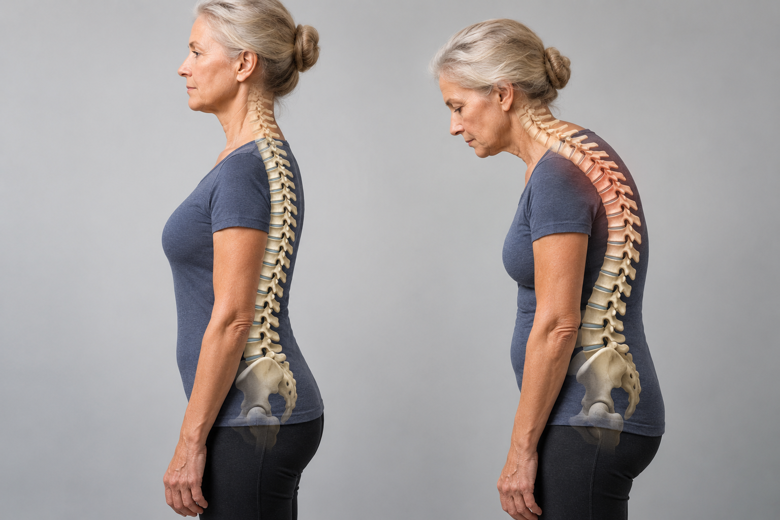
Age-related hyperkyphosis affects approximately 20–40% of older adults and is characterized by an excessive anterior curvature of the thoracic spine, often associated with aging.¹
Definition and Normal Range
A certain degree of anterior curvature in the thoracic spine is normal because of the shape of the vertebral bodies and intervertebral discs.
A kyphosis angle greater than 40° is commonly defined as hyperkyphosis.¹
Causes of Age-Related Hyperkyphosis
General contributing factors include:
Poor posture: Prolonged poor postural control can increase the kyphotic angle.
Dehydration of the intervertebral discs: Reduced fluid content in the discs leads to loss of height and support.
Reduced strength of the spinal extensors: Weakening of the back muscles can contribute to further curvature of the spine.
Consequences of Hyperkyphosis
Age-related hyperkyphosis can lead to several problems affecting physical, psychological, and social health:
Physical symptoms:
Back pain and tenderness.
Neck pain.
Numbness in the upper extremities or buttock region.
Psychological consequences:
Reduced psychosocial well-being, including depression and low self-esteem.²
Functional limitations:
Reduced rib-cage mobility, which may contribute to breathing difficulties.
Increased biomechanical stress on the spine, which may raise the risk of vertebral compression fractures.
Risk of falls and fractures:
Impaired gait and balance can increase the risk of falls and injury.
Reduced physical function:
May affect daily activities and basic functional tasks, resulting in reduced quality of life.³⁴
Mortality:
Hyperkyphosis may be a marker or risk factor associated with premature mortality. Studies have found that increasing kyphosis angle is associated with higher mortality, particularly deaths related to pulmonary problems.⁵
Hyperkyphosis represents a significant health concern in older adults because of both its direct physical consequences and its effects on mental health and quality of life. Understanding its causes, symptoms, and consequences is important for developing targeted preventive and therapeutic strategies.
Clinically Relevant Anatomy: Thoracic Spine
The thoracic spine is an important part of the vertebral column and consists of structures that provide both stability and mobility. Its anatomy plays a key role in the development and consequences of age-related hyperkyphosis.
Key Structures of the Thoracic Spine
Bones:
The thoracic spine consists of 12 thoracic vertebrae (T1–T12).
These vertebrae articulate with the rib cage, providing additional stability and protection.
Joints: the rib cage, providing additional stability and protection.
Joints:
The thoracic spine includes both intervertebral joints and
The thoracic spine includes both intervertebral joints and
The thoracic spine includes both intervertebral and costovertebral joints, allowing limited flexion, extension, and rotation.
Nerves:
Thoracic spinal nerves branch from the spinal cord through the intervertebral foramina and supply the trunk and intercostal muscles.
Ligaments:
Strong ligaments, including the ligamentum flavum, supraspinous ligament, and interspinous ligaments, provide additional support and help limit excessive movement.
Intervertebral discs (IV discs):
The discs in the thoracic spine are thinner and more rigid than those in other spinal regions, resulting in less flexibility.
Muscles:
Muscles such as the erector spinae, transversospinalis and intercostal muscles contribute to stabilization and movement of the thoracic spine.
Thoracic Spine: Stiffness and Function
The thoracic spine is relatively rigid compared with the cervical and lumbar regions, mainly because of:
The attachment of the ribs to the thoracic vertebrae.
Tight ligaments that limit excessive movement.
Thin, relatively rigid intervertebral discs.⁶⁷
Clinical Significance
The unique anatomy of the thoracic spine makes this region susceptible to conditions such as age-related hyperkyphosis. Disc stiffness and the influence of the rib cage contribute to reduced mobility, which can amplify the effects of poor posture and muscular weakness over time. Understanding these structures is important for diagnosis and management of hyperkyphosis.
Epidemiology and Etiology of Age-Related Hyperkyphosis
Epidemiology
Variation in thoracic kyphosis:
Throughout life, the kyphosis angle normally varies around 20–29 degrees.
Prevalence:
20–40% of both men and women may have hyperkyphosis.³
Age-related increase:
After the age of 40, the kyphosis angle tends to increase, generally more rapidly in women than in men.
In women:
The average angle ranges from approximately 43–52 degrees at ages 55–60.
It may increase to around 52 degrees between the ages of 76 and 80.
Etiology
Hyperkyphosis is an excessive thoracic curvature that may develop because of several underlying conditions and contributing factors:
Medical conditions:
Osteoporosis and vertebral fractures:
These are common contributors to hyperkyphosis, although only around one third of people with severe kyphosis have radiographically confirmed vertebral fractures.⁵
Psychosocial factors:
Depression, insecurity, low mood, and anxiety may contribute to poor posture and increased kyphosis.⁸⁹
Changes in connective tissue:
A gradual reduction in connective-tissue elasticity and mobility can reduce the body's ability to resist gravity, contributing to a forward-flexed posture.¹⁰³
Muscle weakness:
Weak spinal extensor muscles reduce the ability to maintain an upright posture.¹⁰
Sensory systems:
Loss of function in the somatosensory, visual, and vestibular systems can impair postural control.
Reduced proprioception and vibration sense from the lower-extremity joints may impair the ability to perceive vertical alignment.³
Hyperkyphosis is a complex condition with multiple contributing factors that can affect both physical and psychosocial health. Understanding its epidemiology and etiology is important for early intervention, prevention, and treatment to reduce functional consequences and improve quality of life.
Characteristics and Clinical Presentation of Thoracic Hyperkyphosis
Thoracic hyperkyphosis is characterized by an increased anterior curvature of the thoracic spine and may develop gradually over time. Patients may not notice the change themselves, while family, friends, or healthcare professionals may identify it. A sudden increase in spinal rounding should be medically assessed because it may be associated with other health problems.
Main Symptoms of Thoracic Hyperkyphosis
Increased anterior curvature of the thoracic spine:
A gradually developing rounded upper back that may become more prominent over time. The changes can be subtle and may not be noticed by the patient but may be visible to others.
Difficulty rising from a chair:
People with hyperkyphosis may have difficulty standing up without using their arms for support. This is often related to reduced strength and flexibility of the spinal extensors and surrounding musculature.
Poor balance:
Many patients experience persistent uncertainty about their balance and may feel at risk of falling. This can be related to reduced proprioception and altered sensory input.
Slower walking:
Walking may become noticeably slower, with reduced step length and gait speed. This may represent a compensation for impaired balance.
Wider base of support during walking and standing:
To reduce fall risk, patients may adopt a wider stance, which can influence both posture and gait pattern.
Reduced stair-climbing speed:
Stair climbing may become more challenging and slower, often because of reduced strength and balance.
Breathing difficulties (severe cases):
In severe cases, hyperkyphosis may contribute to breathing difficulties because of reduced pulmonary vital capacity. This is related to restricted thoracic mobility, which can limit lung expansion.³
Clinical Significance
These symptoms can directly affect functional ability and quality of life. Physical limitations may also contribute to secondary problems such as reduced mobility, social isolation, and loss of independence in daily activities. Accurate assessment of symptoms and functional limitations is therefore important when developing an effective treatment plan.
Differential Diagnoses for Thoracic Hyperkyphosis
Thoracic hyperkyphosis can have several underlying causes, and accurate diagnosis requires consideration of relevant differential diagnoses. These conditions may present similarly but differ in etiology, symptoms, and treatment strategies.
1. Osteoporosis
Description:
A systemic condition characterized by reduced bone mineral density and increased fracture risk, particularly in the thoracic spine.
Relevance:
Osteoporotic compression fractures can lead to a gradual increase in the kyphosis angle.
Diagnosis:
DXA scanning to measure bone mineral density.
Radiography can identify fractures or loss of vertebral body height.
2. Vertebral Fractures
Description:
Compression fractures in the thoracic spine may occur because of trauma, osteoporosis, or pathological weakening of bone.
Relevance:
Fractures may cause acute pain and a noticeable change in spinal curvature.
Diagnosis:
Radiographs may show vertebral collapse or wedge-shaped vertebral bodies.
3. Muscle Imbalance Caused by Neuromuscular Disease
Description:
Conditions affecting muscle strength and control, such as cerebral palsy or muscular dystrophy, can create imbalance between muscles that stabilize and move the spine.
Relevance:
Uneven muscle forces may contribute to increasing thoracic curvature over time.
Diagnosis:
Clinical assessment of neuromuscular function and strength.
Electromyography (EMG) may be used to evaluate muscle function.
4. Ankylosing Spondylitis
Description:
An inflammatory condition primarily affecting the spine that can cause stiffness and kyphotic deformity.
Relevance:
Chronic inflammation may result in progressive spinal fusion and increased kyphosis.
Diagnosis:
Radiography may demonstrate a characteristic 'bamboo spine' appearance.
HLA-B27 testing may support the diagnosis in the appropriate clinical context.
5. Scheuermann's Kyphosis
Description:
A structural deformity of the thoracic spine, usually diagnosed during adolescence.
Relevance:
Characterized by wedging of thoracic vertebrae, resulting in a relatively fixed hyperkyphosis.
Diagnosis:
Radiographic findings typically include three or more adjacent wedged vertebrae with at least 5° of wedging.
Diagnostic Procedures for Measuring Thoracic
Hyperkyphosis
Effective assessment of hyperkyphosis requires precise and reliable measurement tools. Several validated methods are used clinically to evaluate thoracic kyphosis, including radiographic and non-invasive techniques.
1. Radiographic Measurement of the Cobb Angle (Gold Standard)
Description:
Standing lateral radiographs of the thoracic spine are used to measure the Cobb angle.
This method is considered the reference standard for quantifying thoracic kyphosis.
Procedure:
The angle is calculated using lines drawn along the upper endplates of the upper thoracic vertebrae and the lower endplates of the lower thoracic vertebrae, commonly spanning approximately T1/T2 to T11/T12 depending on image visibility.
Advantages:
Highly accurate and well validated for diagnosis.
Limitations:
Requires exposure to ionizing radiation.⁵
2. Non-Invasive Methods
Several radiation-free methods can be used in clinical settings to measure kyphosis:
a) Debrunner Kyphometer
Description:
An instrument with two arms positioned at the upper and lower limits of the thoracic curvature, typically around T2/T3 and T11/T12.
Function:
Measures the kyphosis angle and has shown a strong correlation with radiographic Cobb-angle measurements.
Advantages:
Non-invasive and easy to use in clinical settings.
Good correlation with radiographic measurements.¹¹
b) Flexicurve Ruler
Description:
A flexible ruler molded to the spinal curvature from C7 to approximately L5–S1.
Kyphosis Index:
Calculated as the width divided by the length of the thoracic curve, multiplied by 100.
A value above 13 may indicate hyperkyphosis.¹²
Advantages:
Simple, cost-effective, and suitable for a variety of clinical settings.
c) Spinal Mouse®
Description:
A computer-assisted instrument used to assess spinal curvature without radiation exposure.
Function:
Measures thoracic kyphosis through direct contact with the skin and transfers the data to a computer for analysis.
Advantages:
Non-invasive and safe for repeated measurements.
Disadvantages:
Requires specialized equipment and training.¹³
3. Comparison Between Methods
There is a moderate correlation (ICC = 0.68) between radiographic measurements and standing clinical measurements of kyphosis.¹⁴
Non-invasive methods such as the kyphometer and Spinal Mouse are practical alternatives but may be less accurate than radiographic measurements.
Outcome Measures for Hyperkyphosis: Evaluation of Function and Health
Measuring treatment outcomes in patients with hyperkyphosis is important for assessing functional limitations, pain, and quality of life. Several validated outcome measures are used for this purpose, with specific tools addressing both physical and psychological aspects.
1. Scoliosis Research Society Outcomes Instrument (SRS-22)
Description:
A comprehensive instrument assessing five key domains:
Function.
Pain.
Self-image.
Mental health.
Satisfaction with treatment.
Relevance to hyperkyphosis:
Research shows associations between hyperkyphosis and:
Increased pain.
Lower self-image.
Reduced overall function and activity.
Correlation coefficients for kyphosis (0.40–0.66) are substantially higher than those for scoliosis magnitude versus SRS scores (0.16–0.26), suggesting that SRS-22 may be useful for assessing hyperkyphosis.¹⁵
2. Occiput-to-Wall Distance
Description:
Measures the distance between the occiput and the wall while the patient stands upright with the heels against the wall.
Relevance:
A greater distance indicates increased thoracic kyphosis.
A simple and rapid test that provides an indication of postural change.
3. Tragus-to-Wall Distance
Description:
Measures the distance from the tragus of the ear to the wall while the patient stands upright.
Relevance:
Used as a measure of forward-flexed posture, which is common in people with hyperkyphosis.
4. Neck Pain and Disability Scale (NPAD)
Description:
A scale assessing the severity of neck pain and functional limitations in daily activities.
Relevance:
In people with hyperkyphosis, neck pain may develop as a secondary consequence of postural dysfunction.
5. Timed Up and Go (TUG)
Description:
A functional test in which the patient rises from a chair, walks three meters, turns, returns, and sits down again.
Relevance:
Assesses gait speed, balance, and functional mobility.
Useful for evaluating fall risk, which may be increased in people with hyperkyphosis.
Using outcome measures such as SRS-22, distance measurements such as occiput-to-wall and tragus-to-wall, NPAD, and the TUG provides a broad assessment of the physical and psychological consequences of hyperkyphosis. These tools can help identify areas requiring intervention and monitor progress during treatment. The choice of instrument should be adapted to the patient's needs and the clinical setting.
Clinical Examination of Hyperkyphosis
Hyperkyphotic posture can be assessed clinically using visual and manual methods. Although these methods do not replace radiography as the reference standard for precise angular measurement, they can provide useful information about postural changes and functional limitations.
1. Visual Assessment
Qualitative evaluation:
The therapist assesses the anterior curvature of the thoracic spine.
Changes in thoracic curvature may be secondary to postural compensations in other regions, such as the lumbar spine.
2. Standardized Tests
Tragus-to-wall test:
Measures the distance from the tragus of the ear to the wall while the patient stands upright.
Purpose:
To assess forward-flexed posture and the degree of kyphosis.
Occiput-to-wall test:
Measures the distance between the occiput and the wall while the patient stands with the heels against the wall.
A greater distance indicates more pronounced thoracic kyphosis.
3. Measurement of Head Support Height
1.5 cm blocks:
Measures the number of 1.5 cm blocks required to support the head in a supine position.
Purpose:
Provides a quantitative estimate of head position and support requirements related to kyphotic curvature.
Although these clinical methods cannot replace radiography for precise measurement of kyphosis angles, they provide a rapid and non-invasive assessment that can help identify hyperkyphosis and its effect on posture and function. They are particularly useful in settings where radiography is unavailable or undesirable.
Medical Treatment of Age-Related Hyperkyphosis
Management of age-related hyperkyphosis may include pharmacological and surgical approaches depending on underlying causes such as low bone density or vertebral fractures. Treatment aims to reduce pain, improve function, and limit progression of the condition.
1. Pharmacological Treatment
Goal:
Strengthen bone and reduce the risk of further injury and fractures.
Medication classes:
Antiresorptive agents:
Reduce bone resorption associated with osteoporosis.
Examples include bisphosphonates such as alendronate and zoledronic acid.
Bone-forming medications:
Stimulate new bone formation.
Examples include teriparatide (a PTH analogue) and romosozumab (a sclerostin inhibitor).³
2. Surgical Treatment
For patients with severe symptoms that do not respond to conservative treatment, surgical procedures may be considered. Common options include:
a) Kyphoplasty
Description:
A procedure in which a balloon catheter is inserted into a compressed vertebral body and inflated in an attempt to restore height. The cavity is then filled with acrylic bone cement for stabilization.
Advantages:
May reduce pain.
May improve vertebral deformity.
May reduce the kyphosis angle in selected cases.¹⁷¹⁸
Risks:
Cement leakage can damage surrounding structures, including neural structures such as the spinal cord.
b) Vertebroplasty
Description:
Acrylic bone cement is injected directly into the compressed vertebral body to stabilize the fracture.
Advantages:
May reduce pain and physical disability.
May help prevent further vertebral collapse.¹⁸¹⁹
Risks:
Similar to kyphoplasty, including the risk of cement leakage.
Physiotherapy Treatment
Physiotherapy is an important component of management for age-related hyperkyphosis and focuses on reducing symptoms, improving function, and limiting further progression. Although research suggests that exercise produces a moderate reduction in kyphosis angle, approximately 1.67°–3.74° in some studies, physiotherapy remains important for improving overall function and quality of life.⁴
Goals of Physiotherapy
Increase strength of the spinal extensor muscles.
Improve mobility of the thoracic spine.
Increase postural awareness.
Reduce the risk of vertebral compression fractures.
Maintain or restore physical function and limit further deterioration.³
Treatment Strategies
Physiotherapy should be individualized according to each patient's needs and capacity, with guidance from a qualified physiotherapist. The following strategies may be included in the treatment plan:
1. Postural Correction
Exercise:
Stretching and strengthening exercises aimed at reducing excessive thoracic flexion and improving postural control.
Focus on thoracic extension and activation of the spinal extensor muscles.
Example: exercises emphasizing thoracic extension.
Use of individually fitted braces or therapeutic taping:
May provide temporary support and reduce the apparent curvature angle in selected patients.
2. Breathing Techniques
Purpose:
Improve exercise tolerance by supporting ventilation and respiratory capacity.
Examples:
Diaphragmatic breathing exercises to train respiratory muscles and improve ventilation.
3. Pain Relief
Modalities:
Heat or ice may be used to reduce discomfort.
TENS (transcutaneous electrical nerve stimulation) may be used for pain relief.
4. Myofascial and Soft-Tissue Therapy
Manual techniques:
Myofascial treatment and shoulder mobilization may be used to improve mobility and reduce muscular tension.
5. Education
Purpose:
Increase the patient's understanding of posture, activity modification, and ergonomics in daily life.
Areas of focus:
Appropriate performance of daily activities to reduce unnecessary spinal loading.
6. Balance and Gait Training
Purpose:
Reduce fall risk and improve overall physical capacity.
Exercises:
Static and dynamic balance exercises.
Gait training with attention to posture and step length.
Clinical Significance
Physiotherapy plays a key role in managing age-related hyperkyphosis. Although exercise alone may produce only a moderate reduction in curvature, a combination of postural correction, strengthening, breathing techniques, and education can improve functional capacity and quality of life. Long-term follow-up and individualized exercise are important for maintaining benefits.
Exercise Methods for Managing Hyperkyphosis
Effective management of hyperkyphosis may include a combination of manual mobilization, strengthening, mobility exercises, and postural correction. These approaches have been examined in randomized controlled trials and aim to reduce kyphosis angle, improve function, and enhance quality of life.
1. Hatha Yoga
Modified for hyperkyphosis:
Yoga positions may include:
Breathing techniques: To support respiratory function and promote relaxation.
Stretching tight muscle groups: For example, the pectoral muscles and latissimus dorsi.
Strengthening weak muscle groups: Particularly the rhomboids and other scapular stabilizers.²¹
2. Manual Mobilization
Application:
Mobilizations may be performed according to the Maitland Joint Mobilization Grading Scale.
Treatment may focus on the thoracic spine to improve mobility and reduce stiffness.²²²³
3. Active Mobilization and Stretching
Areas of focus:
Mobilization of the pectoral region, shoulders, and latissimus dorsi.
Improved range of motion and flexibility in shortened or tight muscles.²²²³²⁴
4. Strength Training
Targeted muscle groups:
Thoracic extension: Activation of the paraspinal muscles.
Scapular adductors: Improve scapular stability.
Abdominal muscles: Provide trunk support and postural control.
Shoulder movements: Flexion, adduction, and abduction.
Standing elbow flexion/extension.²²²⁵²⁶²⁷
5. Postural Correction and Awareness Training
Methods:
Visual feedback: Mirrors or photographs.
Auditory feedback: Verbal reminders from the therapist.
Tactile feedback: Taping or therapeutic touch.²²²³²⁵
Steps for postural correction:
Explain the problem:
Inform the patient about factors contributing to the abnormal posture.
Demonstrate correct posture:
Each movement is explained carefully:
Gently draw the navel inward and downward with a light abdominal contraction.
Keep the knees slightly flexed.
Draw the shoulder blades together (scapular retraction).
Lift the chest upward.
Keep the chin slightly tucked.
Practice with the patient:
Begin with detailed instructions and repeat until the patient can achieve the desired posture without guidance.
Use supportive aids:
Taping and reminder timers may be used to support postural awareness throughout the day.
Final Remarks
Hyperkyphosis is common among older adults and is associated with increased risk of fractures, reduced physical performance, poorer health, and reduced quality of life. Early screening and implementation of simple, cost-effective interventions such as physiotherapy, exercise, bracing, and taping may:
Reduce hyperkyphosis.
Improve quality of life.
Reduce the risk of future fractures in both men and women.²⁸⁵
Sources:
Jang et al., H. (2015). Effect of thorax correction exercises on flexed posture and chest function in older women wih age-related hyperkyphosis. J. Phys. Ther. Sci. , 27, 1161-1164.
Jang, H.-J., Hughes, L. C., Oh, D.-W., & Kim, S.-Y. (2019). Effects of Corrective Exercise for Thoracic Hyperkyphosis on Posture, Balance, and Well-Being in Older Women: A Double-Blind, Group-Matched Design. Journal of Geriatric Physical Therapy, 42(3), E17–E27. https://doi.org/10.1519/JPT.0000000000000146
Katzman et al., W. B. (2010). Age-related hyperkyphosis: its causes, management, and consequences. J Orthop Sports Phys Ther , 40, 352-360.
Bansal et al., S. (2014). Exercise for Improving Age-Related Hyperkyphotic Posture: A systematic Review. Arch Phys Med Rehabil , 95 (1), 129-140.
Roghani T, Zavieh MK, Manshadi FD, King N, Katzman W. Age-related hyperkyphosis: update of its potential causes and clinical impacts—narrative review. Aging clinical and experimental research. 2017 Aug;29(4):567-77. Available from:https://www.ncbi.nlm.nih.gov/pmc/articles/PMC5316378/
http://www.spineuniverse.com/conditions/kyphosis/anatomy-kyphosis
Mulligan et al., B. (2015). The Mulligan Concept of Manual Therapy: Textbook of Techniques. Chatswood, NSW, Australia: Elsevier.
Fon et al., G. (1980). Thoracic kyphosis: range in normal subjects. AJR Am J Roentgenol , 134 (5), 979-83.
Lewis et al., J. (2010). Clinical measurement of the thoracic kyphosis. A study of the intra-rater reliability in subjects with and without shoulder pain. BMC Musculoskeletal Disorders , 1 (11), 39.
Hinman et al., M. (2004). Comparison of thoracic kyphosis and postural stiffness in younger and older women. Spine J , 4 (4), 413-7.
Korovessis et al. (2001). Prediction of thoracic kyphosis using the Debrunner kyphometer. J Spinal Disord , 67-72.
Teixeira et al. (2007). Reliability and validity of thoracic kyphosis measurements using the flexicurve method. Brazilian Journal of Physical Therapy , 199-204.
Bowtech Spinal Mouse Available from:https://spinalmouse.ro/en (last accessed 18.4.2020)
Kado et al. (2006). Comparing a supine radiologic versus standing clinical measurement of kyphosis in older women: the Fracture Intervention Trial. Spine , 463-467.
Petcharaporn et al. (2007). The Relationship Between Thoracic Hyperkyphosis and the Scoliosis Research Society Outcomes Instrument. Spine , 226-231.
Kado DM. (2009). The rehabilitation of hyperkyphotic posture in the elderly. Eur J Phys Rehabil Med , 583-93
Goh et al. (2000). A comparison of three methods for measuring thoracic kyphosis: implications for clinical studies. Rheumatology , 310-315.
Pradhan et al., B. (2006). Kyphoplasty reduction of osteoporotic vertebral compression fractures: correction of local kyphosis versus overall sagittal alignment. Spine , 15 (31(4)), 435-41.
Wardlaw et al., D. (2009). Efficacy and safety of balloon kyphoplasty compared with non-surgical care for vertebral compression fracture (FREE): a randomised controlled trial. Lancet , 373 (9668), 1016-24.
http://www.moveforwardpt.com/SymptomsConditionsDetail.aspx?cid=a26d7f6b-85dd-4461-9685-be7a8e5d5725
Greendale et al., G. (2009). Yoga decreases kyphosis in senior women and men with adult-onset hyperkyphosis: results of a randomized controlled trial. J Am Geriatr Soc , 57, 1569-79.
Bautmans et al., I. (2010). Rehabilitation using manual mobilization for thoracic kyphosis in elderly postmenopausal patients with osteoporosis. J Rehabil Med. , 42, 129-135.
Bennell et al., K. (2010). Effects of an exercise and manual therapy program on physical impairments, function and quality-of-life in people with osteoporotic vertebral fracture: a randomised, single-blind controlled pilot trial. BMC Musculoskelet Disord , 11-36.
Abreu et al., D. (2012). The effect of physical exercise on thoracic hyperkyphosis in elderly postmenopausal patients with osteoporosis. Osteoporosis Int. , 23, 120.
Benedetti et al., M. (2008). Effects of an adapted physical activity program in a group of elderly subjects wtih flexed posture: clinical and strumental assessment. J Neuroeng Rehabil , 5, 32.
Itoi et al., E. (1994). Effects of back-strengthening exercise on posture in healthy women 49 to 65 years of age. Mayo Clin Proc , 69, 1054-9.
Katzman WB, Wanek L, Shepherd JA, Sellmeyer DE. Age-related hyperkyphosis: its causes, consequences, and management. journal of orthopaedic & sports physical therapy. 2010 Jun;40(6):352-60. Available from:https://www.ncbi.nlm.nih.gov/pmc/articles/PMC2907357/


