

Ankylosing Spondylitis (Axial Spondyloarthritis)

Axial spondyloarthritis is a seronegative spondyloarthritis that primarily affects the spine and pelvis. The term axial spondyloarthritis has been used since 2009, when the diagnostic criteria from the Assessment of Spondyloarthritis International Society (ASAS) were developed. The ASAS criteria made earlier identification of axial spondyloarthritis possible for the first time through the use of magnetic resonance imaging (MRI)¹.
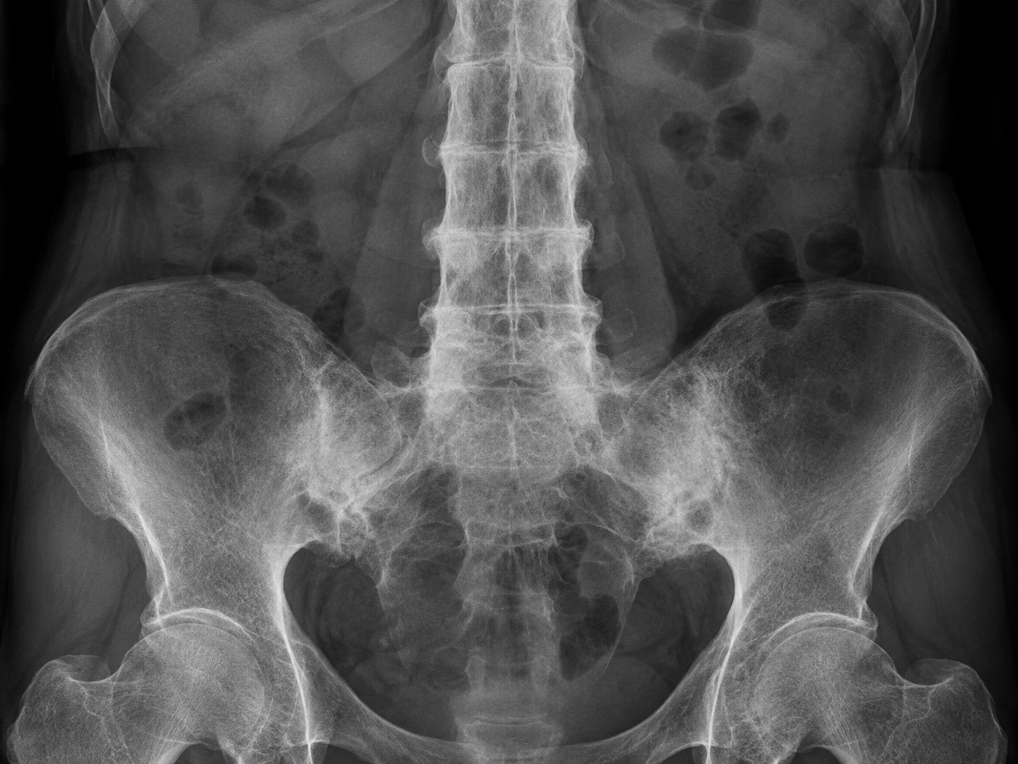
Axial spondyloarthritis is used to describe patients with both non-radiographic and radiographic axial spondyloarthritis. Non-radiographic axial spondyloarthritis does not show signs of disease on X-rays, although changes may be detected on MRI². Radiographic axial spondyloarthritis, also known as ankylosing spondylitis (AS), can be visible on X-rays³.
Axial spondyloarthritis mainly affects the spine, with inflammatory changes leading to pain, stiffness and reduced spinal range of motion⁴⁵. The disease often causes changes in the sacroiliac joints, apophyseal joints, costovertebral joints and intervertebral disc articulations. This results in characteristic inflammatory back pain, which in turn causes structural and functional impairments and reduced quality of life⁶ ⁷ ⁸ ⁹. The affected joints gradually become stiff and sensitive because of new bone formation around joint capsules and cartilage. This reduces range of motion, and in advanced stages the disease can produce the characteristic bamboo-like appearance of the spine, hence the alternative term "bamboo spine".
Although it is not always recognized, axial spondyloarthritis can also cause pain in peripheral joints, particularly the hips, knees, ankles, shoulders and neck. The disease affects synovial and cartilaginous joints as well as the attachment sites of tendons and ligaments⁷⁹. Early diagnosis and treatment are important for controlling pain and stiffness and may help reduce or prevent significant deformity¹⁰.
Clinically Relevant Anatomy
Pain in ankylosing spondylitis (AS) may arise from sacroiliitis, enthesitis and spondylitis[6]. Initially, the sacroiliac joints, located at the base of the spine where the spine connects to the pelvis, become affected. Over time, inflammation spreads to the entheses, which are the areas where tendons and ligaments attach to bone. Eventually, the spine itself is affected by the inflammatory process.
The spine normally consists of 24 vertebrae held together by ligaments and separated by intervertebral discs. In patients with AS, calcium deposits form in the ligaments between and around the intervertebral discs. Accumulation of these deposits leads to ossification[7], beginning at the vertebral margin and extending toward the annulus fibrosus, and is characterized by syndesmophytes[8]. Syndesmophytes are among the most important features of structural spinal damage in ankylosing spondylitis[11]. They are bony outgrowths or projections arising from spinal ligaments where they attach to adjacent vertebral bodies[12]. In very advanced cases, the spine may fuse as a result of new bone formation.
Epidemiology and Etiology
Ankylosing spondylitis (AS) affects between 0.1% and 1.4% of the population[13]. The male-to-female ratio is approximately 2:1 for radiographic axial spondyloarthritis and 1:1 for non-radiographic axial spondyloarthritis[3]. Symptoms typically begin between 20 and 40 years of age[13]. Fewer than 5% of cases begin after the age of 45[13]. AS is more prevalent in Europe (mean 23.8 per 10,000) and Asia (mean 16.7 per 10,000) than in Latin America (mean 10.2 per 10,000)[13].
The etiology of AS is not fully understood, but a strong association between genetic background and environmental factors has been established[14]. Studies have shown that factors such as genetic background, including HLA-B27, microbial infection, endocrine abnormalities and immune-system responses are associated with the development of AS[14]. A direct association between AS and the HLA-B27 antigen has also been demonstrated[14][15]. The exact role of HLA-B27 is unknown, but it is thought to act as a receptor for a triggering antigen that contributes to AS. HLA-B27 is present in 90–95% of patients with ankylosing spondylitis compared with 6–9% of the general population[9]. Environmental factors or bacterial infections may also act as triggers[16].
In the early stages, subchondral bone is infiltrated by granulation tissue, causing small lesions that eventually lead to joint erosion as the adjacent cartilage becomes distorted and the articular surface increasingly irregular[9]. Lesions in the annulus fibrosus eventually undergo ossification, leading to vertebral fusion and the characteristic bamboo-spine appearance. This occurs where the vertebral bodies meet the annular fibers of the intervertebral discs.
When the synovium is affected, it becomes infiltrated by macrophages and lymphocytes. This is followed by replacement of cartilage or fibrous tissue by scar-like fibroblast invasion that rapidly ossifies. The inflammatory response in bone adjacent to involved fibrocartilage, ligaments or periosteum can be marked. When initially affected, the spine may resemble infectious discitis, which can create additional diagnostic confusion for the treating physiotherapist[9].
Clinical Presentation and Features of Inflammatory Back Pain
Inflammatory back pain should be considered when two or more of the following features are present. When four or more features are present, this pattern is considered diagnostic of inflammatory back pain and therefore potentially suggestive of ankylosing spondylitis (AS)[17].
Typical Features
Gradual onset of back pain: Pain that begins around the sacroiliac (SI) joints and buttock region, often presenting as alternating buttock pain, and gradually progresses to involve the entire spine[18].
Age at onset: Symptoms typically begin before the age of 45[17].
Duration longer than 3 months: The pain is persistent rather than transient.
Morning pain and stiffness: Morning stiffness lasting longer than 30 minutes[17].
Night pain: The patient may wake during the second half of the night because of pain, which improves after getting up.
Worsening with inactivity and improvement with movement: Pain and stiffness increase with rest and improve with physical activity[17][19].
The patient is often a young adult, classically a man aged 15–30 years, presenting with gradual onset of low-back pain and stiffness. In the early stages, stiffness is most noticeable after rest and improves with movement. Pain may be worst at night or early in the morning and can wake the patient from sleep.
More specific clinical findings associated with AS include:
Peripheral joint involvement: In addition to the spine, inflammatory arthritis and synovitis may involve peripheral joints such as the hips, shoulders and knees, which are among the most commonly affected limb joints[19].
Eye disease: Ocular involvement occurs in approximately 25% of patients, often as iridocyclitis or conjunctivitis[19].
Chest expansion: Reduced chest expansion and decreased chest-wall movement, commonly less than 2 cm, may be a characteristic sign of AS and can indicate reduced mobility of the axial skeleton[20].
Intermittent low-grade fever, fatigue or weight loss: These systemic symptoms can occur as part of the inflammatory process[21].
Clinical Features to Be Aware Of
Symptoms can vary considerably between patients, and early diagnosis requires attention to both axial and peripheral manifestations. Early recognition and treatment can help prevent more severe deformity and improve quality of life.
Differential Diagnoses for Ankylosing Spondylitis (AS)
When assessing patients with suspected ankylosing spondylitis (AS), several other conditions can produce similar symptoms and should be considered as differential diagnoses. These include:
Common differential diagnoses:
Degenerative Disc Disease: This can cause spinal pain that resembles AS, but degenerative radiographic changes and the absence of inflammatory features help distinguish it from AS.
Herniated Intervertebral Disc: A disc herniation can cause back pain and, in some cases, radiculopathy from nerve compression. Symptoms are generally more localized and often have a different pain pattern.
Fracture and/or dislocation: Acute spinal trauma such as a fracture or dislocation can cause severe back pain and may be confused with early AS symptoms, but there is usually a clear history of trauma.
Osteoarthritis: Primarily a degenerative condition affecting synovial joints. It can cause pain and stiffness, but it does not usually produce the same pattern of axial-skeleton and peripheral-joint inflammation seen in AS.
Spinal Stenosis: Narrowing of the spinal canal can compress neural structures and cause pain and weakness, often related to activity. It can be confused with back pain from AS, but stenosis is typically more closely associated with neurological symptoms.
Spondylolisthesis, Spondylolysis and Spondylosis: These conditions also affect the spine but have different pathophysiology. Spondylolisthesis and spondylolysis often involve vertebral displacement or structural defects, whereas spondylosis describes degenerative changes in spinal joints.
Reactive Arthritis: A type of inflammatory arthritis that often develops after infection, commonly of the urinary or gastrointestinal tract. Reactive arthritis can cause sacroiliac and peripheral-joint pain but differs from AS in etiology and clinical pattern.
Inflammatory Bowel Disease (IBD)-Related Arthritis: Both Crohn's disease and ulcerative colitis may be associated with inflammatory arthritis, particularly involving axial joints. This can resemble AS but occurs in association with underlying intestinal inflammation.
Diffuse Idiopathic Skeletal Hyperostosis (DISH): A condition characterized by excessive bone formation and spinal stiffness. DISH may cause back pain and reduced mobility but lacks the inflammatory features typical of AS.
Rheumatoid Arthritis: An autoimmune disease that primarily affects peripheral joints but can occasionally cause inflammatory changes in the spine. It differs from AS in its systemic inflammatory pattern and characteristic laboratory findings.
Psoriatic Arthritis: An inflammatory arthritis that can involve both axial and peripheral joints, particularly in patients with psoriasis. It can resemble AS, but skin lesions are often also present.
Reiter's Syndrome: A form of reactive arthritis that is often milder and asymmetric, including possible inflammation of the sacroiliac joints. It can be difficult to distinguish from AS, especially in early stages.
All of these conditions should be considered and distinguished from AS to ensure correct diagnosis and treatment. A thorough clinical assessment, including blood tests, imaging such as X-ray or MRI, and the patient's medical history, is essential for establishing an accurate diagnosis.
Diagnostic Procedures for Ankylosing Spondylitis (AS)
The diagnosis of ankylosing spondylitis (AS) is usually based on a combination of thorough subjective and physical examination, laboratory findings and imaging. A comprehensive assessment is necessary because AS shares symptoms with several other conditions.
Common laboratory findings:
HLA-B27 test: This antigen is present in approximately 80–90% of patients with AS, particularly in White and African American populations. However, HLA-B27 may also be present in other inflammatory disorders affecting the bowel or joints[6].
C-reactive protein (CRP): CRP is a marker of inflammation and is often elevated in people with AS, ranging from mildly elevated values above 6 mg/L to substantially higher levels of 20–30 mg/L. Elevated CRP is found in approximately 50–70% of patients with AS[23]. This test can also help identify or exclude other conditions such as spinal infection or cancer.
Erythrocyte sedimentation rate (ESR): Another blood test used to assess inflammation. In AS, approximately one third of patients have an elevated ESR during acute phases of the disease. Other conditions can also cause an elevated ESR[8][23].
Clinical criteria that may assist diagnosis in primary care:
Peripheral manifestations: Features such as dactylitis, enthesitis or peripheral arthritis, including involvement of the plantar fascia or Achilles tendon.
Family history: A first- or second-degree relative with AS or related conditions such as psoriasis, uveitis, reactive arthritis or inflammatory bowel disease may be diagnostically relevant.
Inflammatory back pain: Pain often develops gradually and is characterized by stiffness and pain that are worst in the morning or after rest.
Psoriasis, inflammatory bowel disease or previous episodes of uveitis: A history of these conditions may support a diagnosis within the spondyloarthritis spectrum.
Improvement of back pain after treatment with nonsteroidal anti-inflammatory drugs (NSAIDs): Pain that improves within 24–48 hours of NSAID treatment may support an inflammatory component.
Positive HLA-B27 test: When positive, HLA-B27 can support the diagnosis, particularly when combined with other clinical features[6].
Diagnostic imaging:
Sacroiliitis on X-ray or MRI: X-rays can reveal changes in the sacroiliac joints, which are a characteristic feature of AS. MRI is more sensitive for early signs of sacroiliitis and is often useful for assessing active inflammation[23]
Clinical assessment:
Laboratory tests are often more useful for excluding alternative diagnoses than for confirming AS. Clinical assessment, including the patient's history, symptoms and imaging findings, provides the most accurate diagnostic picture. When AS is suspected, clinicians should also assess disease progression and other associated manifestations.
The diagnosis of AS is therefore based on a combination of clinical observations and specific diagnostic tests and should always be interpreted in the context of the patient's symptoms and overall clinical presentation.
Detection of Ankylosing Spondylitis (AS) with Medical Imaging
Imaging plays an important role in diagnosing ankylosing spondylitis (AS), particularly in confirming disease in later stages. Although X-ray was historically considered the most useful modality for detecting AS, early disease can be difficult to identify. X-rays may remain normal in early stages, whereas established disease can show characteristic radiographic evidence of bone changes and inflammation.
X-rays:
X-rays are effective for identifying changes in later stages of AS, although they are not always reliable early in the disease. In established AS, radiographs may show areas of bone erosion resulting from inflammation and structural damage. X-rays can also show vertebral fusion caused by calcification of the ligaments between vertebrae[23]. Early radiographic findings may include:
Vertebral body squaring
: This occurs following erosion of the anterior corners of the vertebral bodies.Shiny corner sign
: This radiographic sign represents reactive sclerosis secondary to inflammatory erosions at the superior and inferior vertebral endplate corners, known as Romanus lesions[24].MRI (Magnetic Resonance Imaging):
MRI is also used to detect inflammation and sacroiliitis, which are early and important features of AS. MRI is more sensitive than both X-ray and CT for demonstrating sacroiliitis, which often begins with inflammatory changes in the inferior portion of the joint before progressing to bony erosions or sclerosis. MRI may also demonstrate joint-space widening and, eventually, fusion[23].
Characteristic Radiological Signs:
Several radiological signs are characteristic of AS. These include:
Vertebral body squaring
: A change in vertebral shape that can be seen on X-ray.Bamboo spine appearance
: A characteristic appearance produced by vertebral fusion, giving the spine a bamboo-like configuration[23].Dagger sign
: A central dense line on AP radiography of the spine and pelvis caused by ossification of the supraspinous and interspinous ligaments[23].Tram-track sign
: Ossification of the apophyseal joint capsules producing two vertical radiodense lines on either side of the central line on frontal imaging.: Ossification of the apophyseal joint capsules forming two vertical radiodense lines on either side of the central line on frontal imaging.
Trolley-track sign: A combination of the central dagger sign and peripheral tram-track-like lines.
Hatchet deformity: A specific deformity that may be observed in the sacroiliac region[23].
Subchondral erosions, sclerosis and proliferation on the iliac side of the SI joints.
Bridging or fusion of the pubic symphysis: A change that can be seen on radiographs and may indicate progression toward ankylosis[23].
Modified New York Classification Criteria (mNYCC):
The modified New York classification criteria (mNYCC) have historically been used to diagnose axial spondyloarthritis, including AS. The criteria combine clinical findings with radiographic changes to assess disease. Radiographic findings are graded from 0 to 4, where 0 represents normal findings and 4 represents complete ankylosis[6].
Radiographic Criteria:
A definitive diagnosis can be established with the following radiographic combinations[27]:
Grade 3 or 4 changes in both SI joints on X-ray together with at least one clinical criterion.
Grade 3 or 4 changes in one SI joint, or grade 2 changes in both, together with two clinical criteria.
Clinical Diagnostic Criteria:
The patient must also meet at least one of the following clinical criteria:
Lumbar pain and stiffness lasting at least 3 months, improving with exercise but not relieved by rest.
Limited mobility of the lumbar spine in the sagittal and frontal planes.
Reduced chest expansion relative to normal values adjusted for age and sex.
Diagnostic Grading:
Definite AS: A patient can be classified as having definite AS when at least one clinical criterion and the radiographic criterion are fulfilled.
Probable AS: AS may be classified as probable when three clinical criteria are present, or when the radiographic criterion is present without other symptoms fulfilling the clinical criteria.
Both clinical observations and imaging are required for a reliable diagnosis of AS. Combining radiographic findings with clinical symptoms provides a more accurate assessment of the presence and severity of disease.
Ankylosing Spondylitis and Axial Spondyloarthritis
Ankylosing spondylitis (AS) and axial spondyloarthritis are closely related conditions, but they differ in both diagnostic criteria and disease course. Understanding these differences requires consideration of how they are defined and diagnosed, as well as how modern imaging has changed our understanding of these conditions.
Modified New York Criteria and Their Limitations:
The modified New York criteria (mNYCC), developed in 1984, were considered a useful tool for diagnosing AS. They focus on radiographic findings, particularly in the sacroiliac joints, together with clinical symptoms such as low-back pain and stiffness. However, although relatively specific, the criteria are not very sensitive in early disease. This creates challenges in identifying patients during early stages of AS before radiographic changes have developed.
There is also considerable intraobserver and interobserver variability in diagnosis based on X-rays. This highlights the need for more precise diagnostic methods that do not depend solely on conventional radiographs.
The Role of Imaging in Diagnosis:
X-rays may be sufficient for identifying advanced disease progression, including vertebral fusion and severe sacroiliitis. However, early signs of AS are often not visible on conventional radiographs, particularly in the sacroiliac joints. MRI is a more sensitive tool in this setting. MRI can detect sacroiliitis and spinal inflammation in patients with symptoms of AS even when these changes are not visible on X-ray. The ability to detect early inflammatory changes has been central to the broader concept of axial spondyloarthritis (AxSpA).
Axial Spondyloarthritis:
With the development of MRI, it became clear that a substantial proportion of patients with symptoms suggestive of AS but without advanced radiographic sacroiliitis actually had early inflammatory changes in the sacroiliac joints. This led to introduction of the broader diagnosis axial spondyloarthritis (AxSpA), which includes patients with early signs of AS as well as patients with sacroiliitis on MRI but without classic radiographic findings.
ASAS Criteria for Axial Spondyloarthritis:
In 2009, the Assessment of SpondyloArthritis International Society (ASAS) developed classification criteria for axial spondyloarthritis based on imaging, clinical features and laboratory findings. To classify axial spondyloarthritis using the ASAS criteria, the patient must meet the following:
Back pain for at least 3 months beginning before the age of 45.
Sacroiliitis confirmed by MRI or conventional radiography.
At least one clinical or laboratory feature characteristic of spondyloarthritis. This may include findings such as reduced chest expansion, limited spinal mobility or a positive HLA-B27 test.
Alternatively, in people with a relevant clinical history who meet symptom criteria, axial spondyloarthritis can be classified when HLA-B27 is positive together with two features characteristic of spondyloarthritis identified clinically or through laboratory testing.
Subcategories of Axial Spondyloarthritis:
The ASAS criteria divide axial spondyloarthritis into two main groups:
Non-radiographic axial spondyloarthritis (nr-axSpA): This group includes patients with symptoms of spondyloarthritis who do not show classic radiographic signs of sacroiliitis. MRI may demonstrate early disease changes while X-rays remain normal.
Classic ankylosing spondylitis (radiographic axial spondyloarthritis): This refers to patients with classic radiographic signs of AS, such as sacroiliitis and vertebral fusion or bamboo spine, together with clinical symptoms.
Outcome Measures for Ankylosing Spondylitis (AS) and Axial Spondyloarthritis
Effective assessment tools are essential for monitoring disease progression, treatment response and functional status in ankylosing spondylitis (AS) and axial spondyloarthritis. Common outcome measures and assessment tools include:
1. Schober Test
The Schober test is a simple clinical test used to assess lumbar-spine mobility. It measures the change in distance between two skin landmarks over the lumbar spine during forward flexion. Reduced excursion may indicate spinal stiffness, which is common in AS.
2. Occiput to Wall Distance (Fleche Test)
This test assesses the distance between the occiput and the wall while the patient stands upright. The distance provides an indication of thoracic or cervical kyphotic deformity, which can occur in AS.
3. Oswestry Disability Index (ODI)
The ODI is a self-reported questionnaire assessing functional limitations in people with back pain. It evaluates how pain affects daily activities such as walking, standing, sitting, sleeping and work. Higher scores indicate greater disability.
4. Neck Disability Index (NDI)
The NDI is a modified version of the ODI specifically designed to assess functional limitations caused by neck pain. It includes activities such as driving, reading and household tasks.
5. Visual Analogue Scale (VAS)
The VAS is used to assess pain intensity. The patient marks a point on a line ranging from "no pain" to "worst imaginable pain". It provides a subjective measure that can be used to monitor disease development and treatment response.
6. Patient-Specific Functional Scale (PSFS)
The PSFS is another self-reported scale focusing on specific functional problems experienced by the patient. The patient identifies important limitations in daily life and rates how these have been affected by the condition, providing an individualized functional assessment.
7. Bath Ankylosing Spondylitis Metrology Index (BASMI)
BASMI is a comprehensive index assessing physical function and range of motion across several joints and regions of the spine. It includes measures of spinal mobility and hip mobility and is useful for tracking physical status over time[28][10].
8. Revised Leeds Disability Questionnaire (RLDQ)
The RLDQ is used to assess functional limitations in patients with AS and provides insight into how the disease affects daily activities and quality of life[10].
9. European Quality of Life (EuroQoL)
EuroQoL is a measure of health-related quality of life (HRQoL). It evaluates general health and how disease affects perceived quality of life, including pain, mobility and social function[10].
10. Bath AS Disease Activity Index (BASDAI; 0–100)
BASDAI measures disease activity in ankylosing spondylitis. It assesses physical symptoms such as pain, stiffness and fatigue together with the patient's subjective experience of disease. Higher scores indicate greater disease activity[28][10].
11. AMOR Criteria for Spondyloarthritis
The AMOR criteria are used to classify spondyloarthritis based on clinical and radiological findings. The classification includes both axial and peripheral spondyloarthritis and considers symptoms such as back pain, peripheral arthritis and radiographic sacroiliac changes[28].
12. BASFI Index (Bath Ankylosing Spondylitis Functional Index)
BASFI assesses functional problems related to ankylosing spondylitis, including mobility, physical function and ability to perform daily activities. It provides an indication of how the disease affects lifestyle and work capacity[28].
13. BAS-G Index (Bath Ankylosing Spondylitis Global Index)
BAS-G assesses the patient's overall health and well-being in relation to AS and provides an important patient-reported measure of disease burden and quality of life[28].
14. Laboratory Values: C-Reactive Protein (CRP)
CRP is an inflammatory marker used to monitor disease activity and treatment response. Elevated CRP may indicate active systemic inflammation and can help assess response to medical treatment for spondyloarthritis.
Examination in Ankylosing Spondylitis (AS)
A thorough physical examination, particularly of the musculoskeletal system, is important for diagnosing ankylosing spondylitis (AS), especially during early stages when clinical signs may be subtle. Key elements of examination include:
1. Examination of the Sacroiliac Joints and Spine, Including the Neck
In AS, inflammation often begins in the sacroiliac joints and may spread to the spine. Examination of these regions is therefore essential. Specific movements and palpation of the sacroiliac joints can help identify pain or tenderness suggestive of inflammation.
2. Measurement of Chest Expansion and Range of Motion in the Hip and Shoulder Joints
Chest expansion during breathing and range of motion in the hips and shoulders may be affected by AS. Measuring chest expansion helps assess thoracic mobility, which can be reduced when the disease affects the ribs and spine.
3. Enthesitis
Enthesitis, or inflammation where tendons and ligaments attach to bone, is another important feature to assess. Sites such as the heels, knees and hips are commonly affected and can be examined with palpation.
Special Tests for Sacroiliac Joint Involvement:
Sacroiliac Joint Compression Test: This test applies direct side-to-side compression to the pelvis while the patient lies supine. If the sacroiliac joints are inflamed, the maneuver may reproduce pain. Compression can also be applied with the patient side-lying.
Gaenslen's Test: In this test, the patient lies supine while the opposite hip and knee are flexed to stabilize the pelvis. The hip on the tested side is extended, stressing the sacroiliac joint. If the joint is inflamed, the maneuver may reproduce pain.
Straight Leg Raising Test: The patient is asked to raise the leg while keeping the knee extended. Pain during the movement may suggest involvement of the sacroiliac region.
Movement Tests:
Schober Test:
The Schober test assesses lumbar mobility. The examiner marks two points over the lower back and asks the patient to flex forward. Reduced increase in the distance between the points indicates restricted lumbar mobility, which is common in AS.
Lumbar Side-Bending Test:
This test measures lateral flexion of the lumbar spine and assesses range of motion. Restricted side bending may indicate spinal stiffness characteristic of AS.
Chest Expansion Test:
This test measures expansion of the chest during deep inspiration and expiration. Reduced chest expansion may indicate involvement of the thoracic spine and ribs, which becomes more common as AS progresses.
Tragus to Wall Test:
This test assesses neck mobility. The patient stands with the back against a wall, and the distance between the tragus and the wall is measured. Limited cervical mobility is a common feature of AS.
When performed systematically, these physical examinations and special tests help clinicians identify AS in early stages, monitor disease progression and assess response to treatment.
Complications of Ankylosing Spondylitis (AS)
Ankylosing spondylitis (AS) is a chronic inflammatory disease that can lead to several serious complications. These complications may affect multiple organ systems and can substantially influence quality of life. Important complications include:
1. Fractures Related to Osteoporosis
Osteoporosis is common in people with AS and makes bone weaker and more fragile. This increases fracture risk even after relatively minor trauma. Vertebral compression fractures are common and may cause pain and reduced function. As the spine becomes increasingly rigid with disease progression, fractures can also be more difficult to detect in their early stages.
2. Uveitis
Uveitis, or inflammation within the eye, is another important complication of AS. It can cause eye pain, redness and reduced vision and often requires prompt medical treatment to prevent serious ocular damage.
3. Reduced Flexibility
One of the most prominent complications of AS is a substantial reduction in flexibility, particularly in the spine. As inflammation contributes to vertebral fusion, the spine becomes progressively stiffer, which can result in postural changes and reduced range of motion in the neck and back. This can affect the patient's ability to perform daily activities.
4. Restrictions in Lung Function
AS can also affect respiratory function by limiting expansion of the rib cage, particularly when the disease involves the costovertebral joints and thoracic spine. Reduced chest expansion may lead to decreased lung capacity and shortness of breath and, in severe cases, contribute to respiratory impairment.
5. Increased Risk of Cardiovascular Disease
People with AS have an increased risk of cardiovascular disease. Reported complications include aortic insufficiency, aortitis, angina, pericarditis and cardiac conduction abnormalities. The systemic inflammatory process affecting the joints may also involve the cardiovascular system and contribute to significant cardiac disease over time.
6. Fatigue
Fatigue is a common and challenging symptom in people with AS. Chronic inflammation and persistent pain can cause ongoing tiredness that is not necessarily relieved by sleep. Fatigue can substantially reduce quality of life and limit participation in everyday activities.
7. Gastrointestinal Disorders
Gastrointestinal problems, including inflammatory bowel diseases such as Crohn's disease and ulcerative colitis, occur more commonly in people with AS. Symptoms may include diarrhea and abdominal pain and, in some cases, more serious gastrointestinal complications.
AS can cause complications across several organ systems, but appropriate treatment and follow-up can help manage many of these problems effectively. Regular assessment and proactive management are important for preventing or reducing the severity of complications.
Medical Treatment of Ankylosing Spondylitis (AS)
Medical treatment of ankylosing spondylitis (AS) is primarily directed toward controlling symptoms, reducing inflammation and preventing functional decline. The treatment plan varies according to disease severity and treatment response and commonly combines medication with physiotherapy.
First-Line Treatment: NSAIDs (Nonsteroidal Anti-Inflammatory Drugs)
NSAIDs are commonly used to reduce pain and stiffness in the spine and other joints. Although these medications can provide substantial symptomatic relief, they do not directly eliminate the underlying cause of disease.[7] Common NSAIDs used in AS include:
Tolmetin
Sulindac
Naproxen
Diclofenac
Indomethacin (described as particularly effective)[9]
Second-Line Treatment: Corticosteroids and DMARDs
When patients do not respond adequately to NSAIDs, corticosteroid injections or disease-modifying antirheumatic drugs (DMARDs) may be considered.[7] Common DMARDs used in AS include:
Methotrexate
Sulfasalazine
It is important to note that corticosteroids have not demonstrated clear long-term benefit for axial disease, and conventional DMARDs have mainly shown benefit for peripheral-joint symptoms rather than spinal pain or sacroiliitis.[9]
Biologic Medications
Biologic therapy has transformed management of AS, particularly for patients with severe symptoms who do not respond adequately to other medications. Biologic agents target specific molecules involved in the inflammatory process.
Tumor Necrosis Factor Inhibitors (Anti-TNF-α Therapy)
An important component of treatment for AS is the use of TNF inhibitors. Research has shown increased levels of the pro-inflammatory cytokine tumor necrosis factor (TNF) in the sacroiliac joints of patients with ankylosing spondylitis. By inhibiting TNF, these drugs can reduce inflammation and improve symptoms. Well-known TNF inhibitors used in AS include:
Etanercept [29]
Infliximab [22]
Studies have shown that TNF inhibitors can improve symptoms in patients with AS, including pain, stiffness and physical function.
Bisphosphonates
Bisphosphonates affect bone turnover and can reduce bone resorption, which is relevant in AS because the disease involves both bone resorption and new bone formation. Bisphosphonates that have been investigated in AS include:
Neridronate
Pamidronate
These amino-bisphosphonates have been studied for potential anti-inflammatory and bone-related effects in AS.[23]
Adverse Effects and Precautions
All medications, including NSAIDs, corticosteroids, DMARDs and biologic drugs, can cause adverse effects. Patients with other health problems, such as kidney disease or active infection, require careful assessment before treatment. Regular monitoring and close medical follow-up are necessary to identify adverse effects and adjust treatment when required.
Treatment of AS is complex and should be individualized according to the patient's symptoms, disease activity and response to therapy. A combination of medication, physiotherapy and lifestyle measures is often required for optimal outcomes.
Physiotherapy Management of Ankylosing Spondylitis (AS)
Physiotherapy is an important part of ankylosing spondylitis (AS) management and aims to reduce pain, improve spinal mobility and functional capacity, reduce morning stiffness, address postural deformity and support psychosocial well-being.[7] Physiotherapy may also improve quality of life and help maintain function. A Cochrane review by Dagfinrud and colleagues concluded that an individualized, supervised home-exercise program produces better outcomes than no intervention.[10] Core rehabilitation goals include education, an individualized exercise program and physical activity performed at home or in a group setting.[10]
Education
Education plays a central role in physiotherapy management of AS. A study by Sweeney and colleagues suggested that long-term home-based self-management supported by education can be effective.[10] Insufficient education and information may reduce adherence to rehabilitation programs and limit treatment effectiveness.[10] Educational strategies can include written materials, videos, reminder notes and progress charts. Patient motivation and active participation are essential and may be monitored using patient diaries or physiotherapy records.[10]
Treatment Phases in AS
Management of AS can be divided into several phases with different goals and treatment strategies:
Acute Phase (Initial Phase)
During the acute phase, patients commonly experience pain and morning stiffness in the spine.[10]
Primary goal:
Control diffuse and intense pain related to joint stiffness.[10]
Restore proprioception and joint stability, normalize postural patterns and reduce pain.[10]
Pain-relieving interventions combined with pharmacological treatment may help prevent compensatory postures.[10]
Secondary goal:
Preserve flexibility of the pelvis and spine to support effective respiratory function.[10]
Remission Phase
The remission phase may be characterized by persistent pain and restricted mobility in the spine and, in some patients, peripheral joints.[10]
Primary goal:
Goals can be divided into short-term and long-term objectives.[10]
Short-term goals include improving body function and reducing pain.[10]
Long-term goals include pain control, improved activities of daily living, overall function and quality of life.[10]
Chronic Phase
In the chronic phase, diffuse ankylosis may develop, resulting in marked spinal stiffness and a flexed upright posture.[10]
Primary goal:
Help the patient maintain remaining function and achieve the greatest possible degree of independence.
Education about the importance of physiotherapy and body awareness is particularly important in this phase.
Clinical treatment sessions including manual therapy may support adherence to the home-exercise program.[10]
Importance of Physiotherapy Across All Phases
Physiotherapy in AS is not limited to pain reduction; it also aims to preserve functional capacity and quality of life. As the disease progresses and postural changes develop, physiotherapy can help patients maintain independence. Through a structured treatment approach combining physical activity and education, patients can develop more effective self-management strategies.
Important Considerations in Physiotherapy Management
Physiotherapy is an important component of treatment in ankylosing spondylitis (AS), and management should be adapted to the patient's individual needs. Treatment should address both physical symptoms such as pain and stiffness and functional challenges in everyday life.
Fatigue
Fatigue is common in rheumatic diseases such as AS and can substantially affect quality of life. Management strategies may include:
Taking regular short breaks to avoid excessive fatigue.
Avoiding prolonged sitting in the same position.
Maintaining a level of physical activity appropriate to the patient's capacity.
Promoting good sleep hygiene to support recovery and energy levels.
Management of Disease Flares
Patients with AS may experience periods of increased disease activity, often called flares, lasting from days to several weeks. During these episodes, helpful measures may include:
Gentle stretching exercises that maintain mobility without excessively loading the body.
Frequent rest breaks to avoid unnecessary joint stress.
Medication prescribed by the treating physician to reduce inflammation and pain.
Joint Protection and Ergonomics
Joint protection is important for preserving functional ability. Ergonomic principles can be incorporated into activities of daily living (ADLs) by adapting working positions and tasks to reduce unnecessary stress on joints and prevent avoidable strain.
Adherence to Treatment
To achieve the greatest benefit from physiotherapy, it is important to identify factors that may reduce adherence to the treatment plan. These can include:
Patient preferences regarding exercise type, which can be accommodated to reduce boredom and support motivation.
Group therapy may increase motivation and provide social support.
Understanding barriers to adherence is essential, and treatment should be adapted accordingly.
Education and Patient Self-Management
An important component of physiotherapy is education about the disease and how the patient can manage it independently. A strong therapeutic relationship may also improve self-management. Education should explain how AS affects the body, which exercises may be useful and how lifestyle and daily activities can be adapted.
Adaptation of ADL Activities
In addition to physiotherapy exercises, assistive devices and alternative techniques may make daily activities easier. Examples include:
Managing footwear: Slip-on shoes may be easier to use, or the patient may elevate the leg onto a chair or place the foot over the opposite knee to make footwear easier to put on.
Driving: Neck pain and stiffness can make driving difficult. Recommendations may include taking breaks on long journeys and using a small cushion behind the back or under the seat to support better posture.
Clinical Conclusions
Axial spondyloarthritis is a chronic inflammatory joint disease whose exact cause remains incompletely understood. The disease can cause stiffness and pain because of inflammation and new bone formation around joints and entheses. This may lead to structural and functional impairment and reduced quality of life. The axial skeleton and sacroiliac joints are most commonly affected. A combination of medication, including NSAIDs and biologic drugs, together with physiotherapy is commonly recommended. Physiotherapy focuses primarily on mobility exercises, respiratory exercises and postural management to preserve function and quality of life.
Sources:
Dubreuil M, Deodhar A. Axial Spondyloarthritis Classification Criteria–The debate continues. Current opinion in rheumatology. 2017 Jul;29(4):317.
Martey C. Overview of Spondyloarthropathies Course. Plus2020.
Sieper J, Poddubnyy D. Axial spondyloarthritis. The Lancet. 2017 Jul 1;390(10089):73-84.
Martey C. Overview of Spondyloarthropathies Course. Plus2020.
Wenker KJ, Quint JM. Ankylosing Spondylitis. [Updated 2020 Jul 13]. In: StatPearls [Internet]. Treasure Island (FL): StatPearls Publishing; 2020 Jan-. Available from: https://www.ncbi.nlm.nih.gov/books/NBK470173/
Laura A, Haftel H. Shoulder, Knee, and Hip Pain as Initial Symptoms of Juvenile Ankylosing Spondylitis: A Case Report, Journal of Orthopaedic & Sports Physical Therapy [Internet], 1998Feb [cited 2020 Oct 4];27(2):167–172. Available from https://www.jospt.org/doi/10.2519/jospt.1998.27.2.167
Giles L, Singer K. The clinical anatomy and management of back pain series, Volume 2 Clinical Anatomy and Management of Thoracic Spine Pain, Butterworth Heinemann, 2000: 61-66.
Mills K, Page G, Siwek R. A color atlas of low back pain. London: Wolfe Medical Publications, 1990.
Andersson GB, McNeill TW. Lumbar spine syndromes: Evaluation and treatment. Springer Science & Business Media; 2012 Dec 6.
Nava T. Physiotherapy rehabilitation in patients with ankylosing spondylitis. Beyond Rheumatol [Internet]. 2019 Dec 20 [cited 2020 Sep 26];1(2):37-6. Available from: http://beyond-rheumatology.org/index.php/br/article/view/6
Tan S, Wang R, Ward MM. Syndesmophyte growth in ankylosing spondylitis. Current opinion in rheumatology. 2015 Jul;27(4):326.
Syndesmophytes. National Library of Medicine
Dean L, Jones G, MacDonald A, Downham C, Sturrock R, Macfarlane G. Global prevalence of ankylosing spondylitis. Rheumatology [Internet]. 2014 Apr [cited 2020 Sep 26];53(4):650–657. Available from https://academic.oup.com/rheumatology/doi.org/10.1093/rheumatology/ket387
Watad A, Bridgewood C, Russell T, Marzo-Ortega H, Cuthbert R, McGonagle D. The early phases of ankylosing spondylitis: emerging insights from clinical and basic science. Frontiers in immunology. 2018:2668.
Alvarez I, López de Castro JA. HLA-B27 and immunogenetics of spondyloarthropathies. Curr Opin Rheumatol. 2000;12(4):248-253 Alvarez I, de Castro JA. HLA-B27 and immunogenetics of spondyloarthropathies. Current opinion in rheumatology. 2000 Jul 1;12(4):248-53.
Braggins S. Back care: a clinical approach. Churchill Livingstone; 2000.
Taurog JD, Chhabra A, Colbert RA. Ankylosing spondylitis and axial spondyloarthritis. New England Journal of Medicine. 2016 Jun 30;374(26):2563-74.
Baaj A, Praveen V, Mummaneni S, Uribe R, Vaccaro S, Mark S. Handbook of spine surgery. New York: Thieme, 2010:180-182.
Andersson GB, McNeill TW. Lumbar spine syndromes: Evaluation and treatment. Springer Science & Business Media; 2012 Dec 6.
Kataria RK, Brent LH. Spondyloarthropathies. American family physician. 2004 Jun 15;69(12):2853-60.
Goodman Catherine C, Snyder TE. Differential diagnosis for physical therapists: screening for referral. St. Louis: Saunders Elsevier. 2007.
Baaj A, Praveen V, Mummaneni S, Uribe R, Vaccaro S, Mark S. Handbook of spine surgery. New York: Thieme, 2010:180-182.
Mandl P, Navarro-Compán V, Terslev L, Aegerter P, Van Der Heijde D, D'Agostino MA, Baraliakos X, Pedersen SJ, Jurik AG, Naredo E, Schueller-Weidekamm C. EULAR recommendations for the use of imaging in the diagnosis and management of spondyloarthritis in clinical practice. Annals of the rheumatic diseases. 2015 Jul 1;74(7):1327-39.
Shiny corner sign (ankylosing spondylitis). Available from: https://radiopaedia.org/articles/shiny-corner-sign-ankylosing-spondylitis#:~:text=The%20shiny%20corner%20sign%20is,are%20known%20as%20Romanus%20lesions.
Slobodin G, Eshed I. Non-radiographic axial spondyloarthritis. Isr Med Assoc J. 2015 Dec 1;17(12):770-6.
Lambrecht V, Vanhoenacker FM, Van Dyck P, Gielen J, Parizel PM. Ankylosing spondylitis: what remains of the standard radiography anno 2004. JBR-BTR. 2005;88(1):25-30.
Linden SV, Valkenburg HA, Cats A. Evaluation of diagnostic criteria for ankylosing spondylitis. Arthritis & Rheumatism. 1984 Apr;27(4):361-8.
Jordan CL, Rhon DI. Differential diagnosis and management of ankylosing spondylitis masked as adhesive capsulitis: a resident's case problem. Journal of Orthopaedic & Sports Physical Therapy. 2012 Oct;42(10):842-52.
Bullough G, Boachie-Adjei O. Atlas of spinal diseases. Hampshire: Gower Medical Publishing, 1987.
bjchealthAU. Modified Schober's Test. Available from: http://www.youtube.com/watch?v=B9RaFB5BwrQ [last accessed 01/12/12]
bjchealthAU. Lumbar Side Flexion Test. Available from: http://www.youtube.com/watch?v=c-IeFZkPEoE [last accessed 01/12/12]
bjchealthAU. Chest Expansion Test. Available from: http://www.youtube.com/watch?v=SumtVr5c1Qg [last accessed 01/12/12]
Physiotutors. Compression Test / Side-lying Compression | Sacroiliac Joint Provocation. Available from youtube.com/watch?v=pWjvrhWMR4w (last accessed on 20/7/2023
Physiotutors. Gaenslen's Test | Sacroiliac Joint Provocation. Available from https://www.youtube.com/watch?v=ED_bYoQk9d4 (last accessed on 20/7/2023
Physiotutors. Straight Leg Raise or Lasègue's Test for Lumbar Radiculopathy. Available from https://www.youtube.com/watch?v=LdAD9GNv8FI (last accessed on 20/7/2023)
Living with a Chronic Disease – The Story of Ankylosing Spondylitis


