


Anterior Interosseous Nerve Syndrome

The anterior interosseous nerve (AIN) is a terminal motor branch of the median nerve. It branches from the median nerve in the proximal forearm, just below the elbow joint, approximately 5–8 cm distal to the lateral epicondyle and 4 cm distal to the medial epicondyle. It then passes between the two heads of the pronator teres muscle and runs deep along the interosseous membrane, parallel to the anterior interosseous artery.
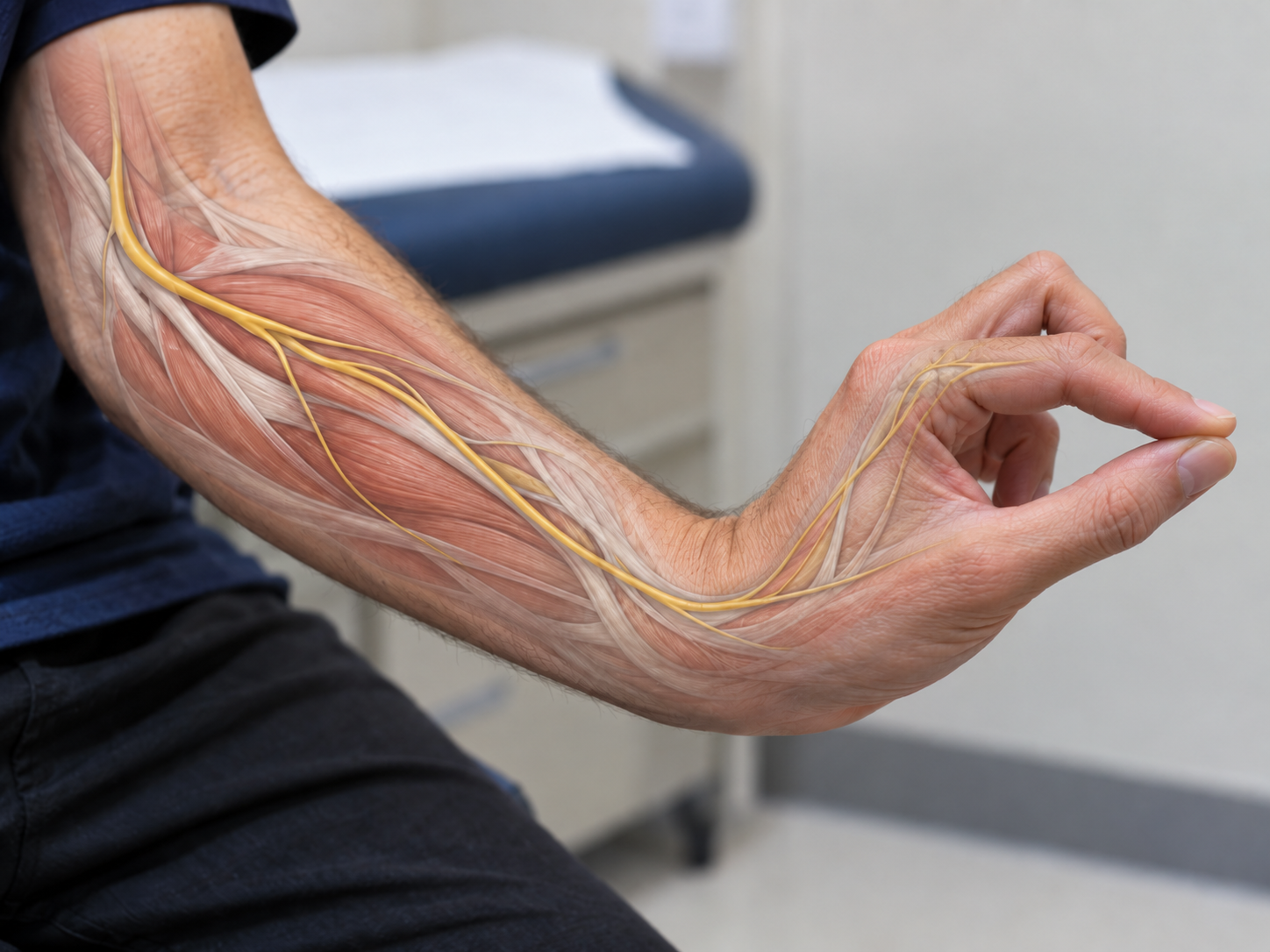
The nerve innervates the following three muscles from proximal to distal:
Flexor pollicis longus (FPL): Responsible for flexion of the interphalangeal joint of the thumb.
Flexor digitorum profundus (FDP): Innervates the radial portion responsible for flexion of the index and middle fingers.
Pronator quadratus (PQ): Produces pronation of the forearm.¹²
Function and Clinical Significance:
The AIN is a purely motor nerve and plays an essential role in precision grip, particularly the pinch movement between the thumb and index finger. An isolated AIN lesion causes weakness in the muscles listed above without sensory deficits. This can result in difficulties with fine motor control and precision grip.¹
Historical Background:
The condition was first described by Parsonage and Turner in 1948, while Leslie Gordon Kiloh and Samuel Nevin in 1952 defined it more specifically as an isolated lesion of the anterior interosseous nerve. This gave rise to the alternative name Kiloh-Nevin syndrome.¹
Clinical Manifestation:
Main symptoms:
Pain: Usually localized to the forearm and often occurs without numbness.
Muscle weakness: Characteristic weakness of the pincer grip between the thumb and index finger.
Motor impairment: Difficulty performing specific tasks requiring precision, such as tying knots or holding small objects.
Causes:
Neuritis: Most cases are thought to occur as a result of transient neuritis.
Compression: May result from anatomical variations such as hypertrophied muscles or fibrous bands.
Trauma: Forearm injuries, often associated with overuse or a direct blow to the region.¹
The purely motor nature of AIN syndrome distinguishes it from other median-nerve-related conditions such as carpal tunnel syndrome, which typically also involves sensory symptoms.
Mechanism of Injury and Pathological Process: Anterior Interosseous Nerve Syndrome
Mechanisms of injury can be divided into spontaneous and traumatic causes:
1. Traumatic Causes:
Forearm fractures: Supracondylar fractures and other forearm fractures may cause compression or injury of the AIN.
Penetrating injuries: Stab wounds and sharp injuries to the forearm region.
Compression from a cast: An improperly fitted or overly tight cast may place pressure on the nerve.
Venipuncture: In rare cases, venipuncture near the median nerve may injure the AIN.
Iatrogenic injury: Injury may occur as a complication following open reduction and internal fixation of fractures (ORIF).
Viral neuritis: A viral infection may cause inflammation of the nerve and affect its function.
2. Spontaneous Causes:
Brachial plexus neuritis: An inflammatory condition affecting nerves of the brachial plexus, including the AIN.
Compartment syndrome: Increased pressure within the forearm compartments may compromise blood supply and nerve function.
Compression neuropathy: The most common site of AIN compression is near the tendinous edge of the deep head of the pronator teres muscle.
Systemic conditions: Rheumatoid arthritis and gout may predispose to nerve compression through inflammation and swelling of adjacent structures.
Prevalence:
AIN syndrome is a rare condition accounting for only 1% of all upper-extremity palsies.¹²
Pathological Process:
Neuropathy: Compression or irritation of the nerve can lead to degeneration of nerve fibers.
Ischemia: Reduced blood supply to the nerve due to compression may damage axons and impair conduction.
Muscle dysfunction: Because the AIN is a purely motor nerve, injury causes weakness or loss of function in the innervated muscles (FPL, FDP and PQ), producing characteristic motor deficits such as a weakened pinch grip.
Clinical Implications:
The most common manifestation of AIN syndrome is weakness of flexion at the interphalangeal joint of the thumb and distal interphalangeal joint of the index finger, which may impair fine motor tasks.
Clinical Presentation of Anterior Interosseous Nerve Syndrome (AIN Syndrome)
The AIN is a purely motor nerve, and true AIN syndrome therefore presents only with motor deficits without sensory symptoms. An overview of the clinical presentation is provided below:
Common symptoms:
Pain: Diffuse, poorly localized pain in the forearm and cubital fossa is often the first symptom.
Motor weakness:
Flexion deficits: The patient has difficulty flexing the interphalangeal joint of the thumb and the distal interphalangeal joint of the index finger.
Grip-function problems: Inability to form an "OK" sign between the thumb and index finger because of weakness in FPL and FDP.
Difficulty with daily activities: The patient may report difficulty making a fist, buttoning shirts or performing fine motor tasks.
Findings on Physical Examination:
Pinch Grip Test: During testing, the patient may be unable to form a normal "OK" sign and instead pinches using more extended thumb and index-finger joints.
Hand of Benediction: When asked to make a fist, the patient may be unable to flex the second and third digits normally, producing a characteristic "hand of benediction." This differs from ulnar claw hand.
No sensory deficits: Sensory complaints are absent, which is useful in differential diagnosis.
Anatomy and Variations Affecting Symptoms:
Martin-Gruber Anastomosis:
Occurs in up to 25% of the population, where branches from the median nerve communicate with the ulnar nerve.
This can produce atypical patterns of motor innervation in the forearm and hand and may mask or alter the classic features of AIN syndrome.
Diagnostic Importance:
Distinguishing AIN syndrome from other neuropathies, including median-nerve lesions at the wrist or more proximally in the arm, is essential.
The pinch-grip test and assessment of motor function are important clinical tools for establishing an accurate diagnosis.
Diagnostic Methods for Anterior Interosseous Nerve Syndrome (AIN Syndrome)
Diagnosis of AIN syndrome requires a combination of electrophysiological studies and diagnostic imaging, which can help identify underlying causes and distinguish AIN syndrome from other neuropathies.
1. Electrophysiological Studies
Nerve conduction studies:
Sensory nerve-conduction studies of the median nerve are expected to be normal, because the AIN has no cutaneous sensory innervation.
Electromyography (EMG):
May show characteristic abnormalities in the following muscles:
Flexor Pollicis Longus (FPL): Reduced function or denervation activity.
Radial portion of Flexor Digitorum Profundus (FDP): Abnormal activity in the portion controlling flexion of the index finger.
Pronator Quadratus (PQ): Reduced or abnormal activity.
EMG can help distinguish AIN syndrome from neuralgic amyotrophy such as Parsonage-Turner syndrome, and from compression neuropathies.
2. Magnetic Resonance Imaging (MRI)
Indications:
Evaluates possible structural causes of nerve compression, including:
Thickening or fibrosis of the pronator teres muscle.
Compression caused by osteophytes or tumors.
Inflammatory or traumatic changes around the median nerve and its branches.
Diagnostic Differentiation
The combination of normal sensory nerve-conduction studies and pathological EMG findings in specific muscles (FPL, FDP and PQ) strongly supports the diagnosis of AIN syndrome.
MRI can help exclude other causes of compression such as tumors, trauma or inflammatory conditions.
Outcome Measures for Anterior Interosseous Nerve Syndrome (AIN Syndrome)
Outcome measures are important for assessing prognosis, setting treatment goals and monitoring progress after AIN syndrome. Relevant tools and methods include:
1. Manual Muscle Test (MMT)
Description:
A simple method used to evaluate strength in the affected muscles, including flexor pollicis longus, flexor digitorum profundus and pronator quadratus.
Strength is graded on a scale from 0 to 5, where:
0: No detectable muscle activity.
5: Full strength against maximal resistance.
Clinical use:
Documents changes in muscle strength over time and provides an objective measure of functional recovery.
2. Nine-Hole Peg Test (NHPT)
Description:
Assesses fine motor skills and hand dexterity by measuring how quickly the patient can place and remove small pegs from holes.
Clinical use:
Can be used to monitor improvement in precision grip and coordination after nerve injury.
3. Disabilities of the Arm, Shoulder, and Hand (DASH)
Description:
A self-reported questionnaire measuring physical function and symptoms related to the upper extremity.
The patient rates limitations in daily activities on a scale from 0 to 100.
Clinical use:
Evaluation of functional limitations and symptom-related improvement.
4. Purdue Pegboard Test
Description:
Assesses finger dexterity by measuring the ability to place pegs into holes as quickly as possible within a specified period.
Clinical use:
Particularly useful for measuring fine motor skills and hand and finger coordination.
5. Grip and Pinch Strength Tests
Description:
Measures grip strength and pinch function using a dynamometer or pinch gauge.
Assesses how effectively the patient can perform tasks requiring thumb-to-index-finger grip.
Clinical use:
Quantifies functional loss and documents progress following treatment.
6. Michigan Hand Outcomes Questionnaire (MHQ)
Description:
A self-reported questionnaire assessing hand function, pain, work-related activities and aesthetics.
Clinical use:
Provides a broader understanding of how AIN syndrome affects the patient's quality of life.
Using these outcome measures helps clinicians assess functional loss, set realistic treatment goals and monitor progress throughout rehabilitation. MMT provides a simple and effective method for evaluating muscle strength, while tools such as DASH and NHPT provide a broader assessment of functional limitations.
Treatment and Intervention for Anterior Interosseous Nerve Syndrome (AIN Syndrome)
A multidisciplinary approach is optimal for AIN syndrome because it allows both primary and secondary contributors to be addressed. Management may include conservative strategies such as rest, physiotherapy and medical treatment, with surgery reserved for cases that do not improve with conservative care.
Conservative Treatment
1. Rest and Activity Modification:
Goal: Reduce loading of the affected nerve.
Patients are advised to avoid activities that aggravate symptoms, including repetitive movements or heavy lifting.
2. Medical Treatment:
Analgesics: For pain relief.
Anti-inflammatory medication (NSAIDs): May be used to reduce inflammation around the median nerve.
Corticosteroid injections: May be considered to reduce localized inflammation.
3. Physiotherapy:
Goal: Improve flexibility, reduce neural irritation and restore motor function.
Exercises may include:
Neural mobilization (nerve gliding): To improve nerve excursion and reduce mechanical sensitivity.
Stretching exercises: Particularly for the flexor musculature of the forearm.
Strength training: Gradually progressed once symptoms improve.
Ergonomic modifications: Adjusting work position or tool use to reduce loading of the arm.
Follow-up of Conservative Treatment
Timeframe: Patients may be treated con
servatively for up to 6 months before surgery is considered.
Evaluation: If the patient does not improve or symptoms worsen, surgical intervention may be considered.
Surgical Treatment
1. Indications for Surgery:
No clinical improvement after approximately 6 months of conservative treatment.
Confirmed compression neuropathy, for example on MRI or electromyography.
Progressive symptoms that substantially impair function.
2. Surgical Procedures:
Neurolysis and decompression: Surgical release of the nerve from compressive structures.
Surgical outcome:
Studies suggest that surgery performed within 8 months of symptom onset may provide a better prognosis than intervention after 12 months.[5]
Prognosis
Good prognosis: Most patients improve with conservative treatment within 6 months.
Surgical intervention: Patients undergoing surgery within an appropriate timeframe may have a high likelihood of improved muscle strength on MMT and better function.
Physiotherapy Treatment
Management of AIN syndrome varies depending on the underlying cause. In spontaneous AIN syndrome, physiotherapy may play an important role, particularly because the precise cause is often uncertain. As a purely motor neuropathy, physiotherapy focuses on the following goals:
Treatment Goals
Maintain muscle integrity: Reduce the risk of muscle atrophy and weakness.
Reduce neural hypersensitivity: Limit excessive neural irritability.
Prevent contractures: Maintain mobility of the joints and muscles.
Increase strength in affected muscles: Improve function of the hand and forearm.
Interventions
Treatment should be individualized and adjusted according to regular reassessment. Recommended interventions may include:
Neural mobilization:
Exercises designed to improve nerve excursion in the upper extremity.
May reduce mechanical sensitivity and support normal nerve movement.
Stretching exercises:
Particularly directed at the flexor musculature of the forearm to improve flexibility.
Electrical stimulation:
TENS (Transcutaneous Electrical Nerve Stimulation): May be used for pain relief and to reduce neural hypersensitivity.
NMES (Neuromuscular Electrical Stimulation): May be used to support muscle activation and strength.
Soft-tissue mobilization:
Massage and mobilization of the forearm musculature may be used to reduce tension and trigger-point sensitivity.
Strength training:
Gradual rebuilding of hand and wrist strength using isometric and isotonic exercises.
Example Treatment Protocol
A case report published in 2016 described a combination of treatment techniques for a patient with AIN syndrome associated with an active trigger point in the middle forearm:
Cryomassage: Used to reduce local irritation and pain.
Neural mobilization: Exercises aimed at promoting normal nerve movement.
TENS: Used for pain relief.
Strain-Counterstrain (SCS): An indirect myofascial technique used to address somatic dysfunction within the neuromuscular system.
Results: After two weeks of treatment, four times per week, the patient demonstrated:
Improved pinch grip with strength increased to eight pounds.
An inactive trigger point on palpation.
Improved handwriting.
Follow-up and Evaluation
Regular follow-up is essential for monitoring progress and adjusting treatment. Monthly assessments may include:
Muscle strength: Assessed with manual muscle testing (MMT).
Pinch grip: Objectively measured with a dynamometer or pinch gauge.
Functional ability: Assessed through the patient's ability to perform daily activities.
Differential Diagnoses
AIN syndrome is a rare and sometimes challenging clinical diagnosis. The following conditions should be considered as differential diagnoses:
Stenosing tenosynovitis:
Inflammation or narrowing of the tendon sheath may cause pain and reduced function that can resemble AIN syndrome.
Flexor-tendon adhesions:
Post-traumatic or postoperative adhesions can restrict flexor-tendon excursion and mimic motor weakness associated with AIN syndrome.
Flexor-tendon rupture:
Injury or degeneration of flexor pollicis longus (FPL) or flexor digitorum profundus (FDP) may cause inability to flex the distal phalanx of the thumb or index finger.
Brachial neuritis (Parsonage-Turner syndrome):
An idiopathic inflammatory neuropathy that may cause pain and weakness in the upper extremity. Brachial neuritis often involves multiple nerves and may include sensory symptoms, which can help distinguish it from isolated AIN syndrome.
Importance of Differential Diagnosis
Accurate differentiation is essential to avoid inappropriate treatment. A detailed clinical examination combined with electrodiagnostic testing, imaging such as MRI and review of the patient's medical history can help distinguish AIN syndrome from other causes of similar symptoms. [1]
Sources:
Akhondi H, Varacallo M. Anterior interosseous syndrome. InStatPearls [Internet] 2019 Jun 4. StatPearls Publishing.
Aljawder A, Faqi MK, Mohamed A, Alkhalifa F. Anterior interosseous nerve syndrome diagnosis and intraoperative findings: A case report. International Journal of Surgery Case Reports. 2016 Jan 1;21:44-7.
Radiopaedia. Anterior interosseous nerve syndrome. from: https://radiopaedia.org/articles/anterior-interosseous-nerve-syndrome-1
Anterior Interosseous Nerve - Everything You Need To Know - Dr. Nabil Ebraheim. Available from:https://www.youtube.com/watch?v=R15I0JzYIDc.
Kodama N, Ando K, Takemura Y, Imai S. Treatment of spontaneous anterior interosseous nerve palsy. Journal of Neurosurgery. 2019 Mar 8;132(4):1243-8.
Goyal M, Goyal K, Narkeesh K, Samuel AJ, Sharma S, Chatterjee S. The strain–Counter strain technique in the management of anterior interosseous nerve syndrome: A case report. Journal of Taibah University Medical Sciences. 2017 Feb 1;12(1):70-4.


