


Avascular Necrosis of the Talus

Avascular necrosis (AVN) of the talus is a condition in which the blood supply to the talus is disrupted, resulting in death of bone tissue. This can cause pain, reduced function, and in severe cases collapse of the talus. Early diagnosis and treatment are important to prevent serious complications such as permanent joint damage and restricted mobility.
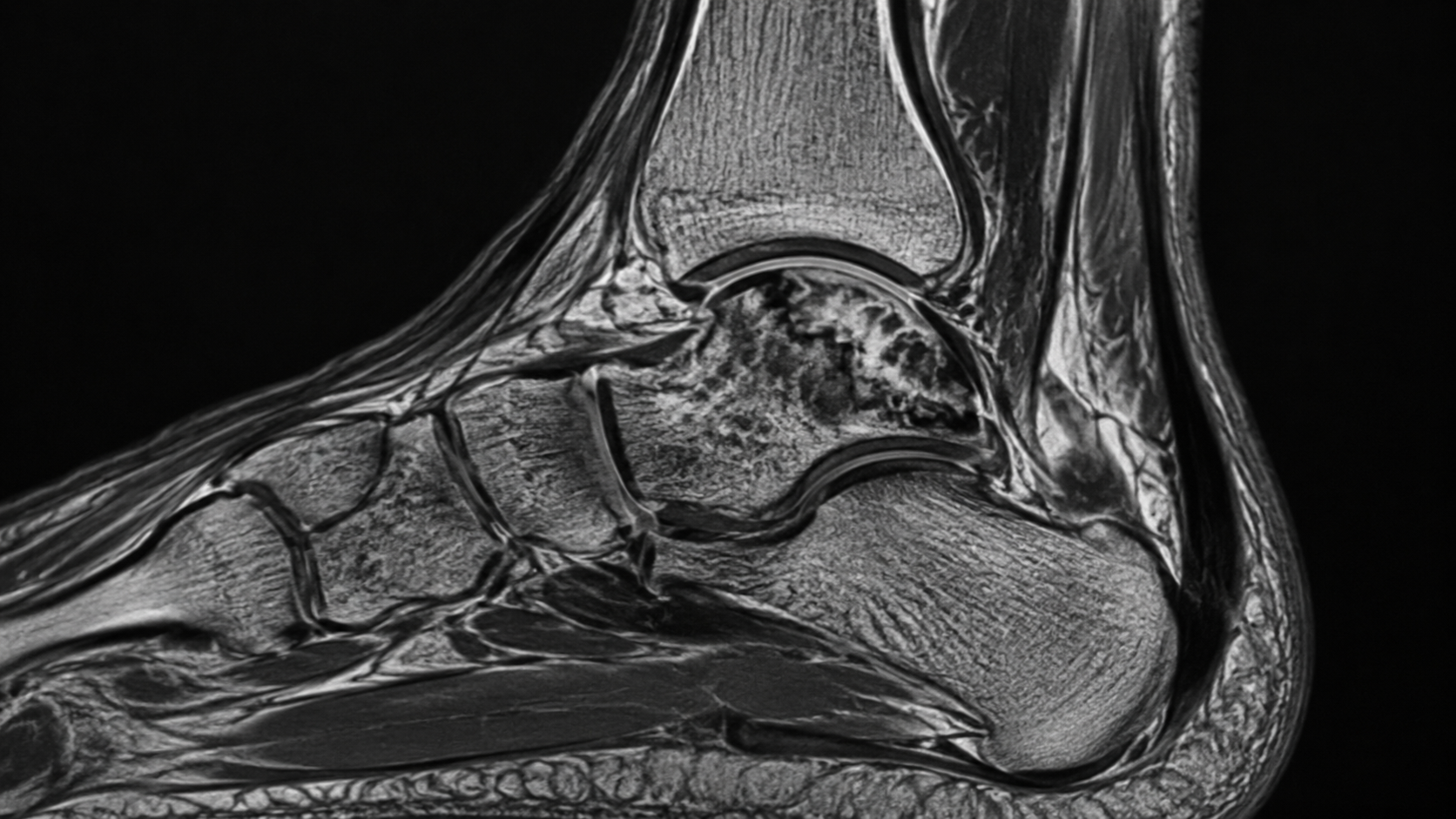
Anatomy of the Talus
Location:
The talus is an important bone in the ankle. It connects the lower-leg bones (tibia and fibula) with the foot and plays a central role in weight-bearing and movement.
Function:
The talus is essential for articulation between the foot and lower leg and enables movements involved in walking, running, and jumping. It also contributes to ankle stability and mobility, and its integrity is important for normal biomechanics.
Causes of Avascular Necrosis of the Talus
AVN of the talus may occur because of several factors that reduce or block its blood supply:
Trauma:
Fractures or dislocations involving the talus can damage the vessels that supply the bone and dramatically increase the risk of AVN, particularly after high-energy injuries such as motor-vehicle accidents or falls from height.[3][4]
Long-term corticosteroid use:
Corticosteroids, often used to treat autoimmune or inflammatory conditions, can impair blood flow to bone, probably through mechanisms affecting fat metabolism or the microcirculation.[5]
Alcohol misuse:
Excessive alcohol consumption may lead to accumulation of fatty substances in blood vessels, reducing blood supply to bone tissue.[6]
Blood disorders:
Conditions such as sickle cell disease can obstruct blood vessels, resulting in reduced oxygen delivery to bone tissue and subsequent cell death.[3]
Decompression sickness:
Rapid changes in pressure, as may occur in divers, can cause gas bubbles to form in the bloodstream and obstruct circulation.
Autoimmune diseases:
Conditions such as lupus can cause inflammation and vascular injury, reducing blood supply to the talus.[3]
Idiopathic causes:
In some cases, the exact cause remains unknown despite extensive investigation.[6]
Importance of Early Intervention
Early diagnosis and treatment of AVN of the talus are essential for preserving bone structure and preventing collapse. Diagnostic imaging such as X-ray and MRI, together with thorough clinical assessment, plays a key role in early detection. Early treatment may slow progression and improve the patient's quality of life.
Symptoms of Avascular Necrosis of the Talus
AVN of the talus usually develops gradually, and symptoms may vary according to disease stage and severity:
Pain:
The most common and often the first symptom. Pain is usually located in the ankle or foot and may begin as mild and intermittent before becoming progressively more intense.
Swelling:
Swelling around the ankle is common, especially in later stages, and may be accompanied by warmth and tenderness.
Restricted range of motion:
The patient may have difficulty moving the ankle freely, which can limit activities such as walking, running, or jumping.
Antalgic gait:
Because of pain and ankle instability, the patient may begin to limp in order to unload the affected area.[7]
Classification of Avascular Necrosis of the Talus
AVN of the talus can be described using the Ficat and Arlet classification, which is used to characterize the progression of osteonecrosis in different bones:
Stage I
Symptoms: Mild pain, often intermittent and without major functional limitation.
Imaging: Plain radiographs appear normal. MRI may, however, demonstrate bone-marrow edema as an early indicator of osteonecrosis.
Stage II
Symptoms: Persistent pain and mild functional impairment.
Imaging: Radiographs show sclerosis or cystic changes. MRI demonstrates more extensive edema and areas of necrotic bone.
Stage III
Symptoms: Severe pain and marked functional impairment affecting daily activities.
Imaging: Radiographs show subchondral fractures, known as the "crescent sign", and possible narrowing of the joint space.
Stage IV
Symptoms: Severe and often disabling pain.
Imaging: Radiographs show collapse of the talus together with advanced arthritic changes, including deformity of the ankle joint.[8]
Importance of Classification
This classification helps clinicians determine disease severity, guides treatment selection, and provides prognostic information. Early intervention in the initial stages may prevent progression to more severe stages in which irreversible damage can occur.
Diagnosis of Avascular Necrosis of the Talus
Physical examination
A thorough physical examination is important for identifying features of AVN, including:
Pain and tenderness: Palpation around the ankle and foot to identify painful areas.
Swelling: Assessment of swelling or redness around the joint.
Range of motion: Testing ankle mobility to identify limitations in dorsiflexion or plantarflexion.
Imaging
Imaging methods are essential for confirming the diagnosis and evaluating severity:
X-rays:
May demonstrate structural changes in the talus, such as sclerosis, cystic changes, or collapse in advanced stages.
Sensitivity is limited in early disease.
MRI:
The gold standard for early diagnosis.
Shows bone-marrow edema and necrotic areas before they become visible on radiographs.
CT scan:
Provides detailed cross-sectional images of bone and is useful for evaluating the complexity of injury or collapse.
Particularly useful for surgical planning.[9]
Treatment of Avascular Necrosis of the Talus
Non-surgical treatment
For patients in early stages or with milder symptoms:
Medication:
Analgesic and anti-inflammatory medications may be used to relieve symptoms.
Bisphosphonates may be considered to reduce bone resorption.
Physiotherapy:
Focuses on maintaining joint function and strength through individualized exercise.
Mobility exercises and strengthening can help prevent muscle atrophy.
Orthotic support:
Braces, rigid soles, or custom footwear may be used to support the foot and reduce loading of the talus.[9][10]
Surgical treatment
In more advanced cases, or when conservative treatment does not provide sufficient benefit:
Core decompression:
Removal or drilling of part of the necrotic bone to reduce intraosseous pressure and promote new blood-vessel formation.
Most effective in earlier stages.
Bone grafting:
Transplantation of healthy bone tissue to the affected area to provide structural support and promote healing.
Arthrodesis:
Fusion of the joint to eliminate pain and stabilize the ankle.
Often used in advanced collapse and severe deformity.
Total ankle replacement:
Replacement of the damaged joint with a prosthesis in very severe cases.
This can preserve movement and reduce pain but requires careful patient selection for optimal outcomes.[9][10]
Prognosis
The outcome of avascular necrosis (AVN) of the talus depends largely on how early the condition is diagnosed and treated. Early detection and intervention can significantly improve prognosis by limiting further joint damage. When diagnosed at an advanced stage, the condition may result in permanent joint damage and disability.[11]
Prevention
The following measures may help reduce the risk of developing AVN of the talus:
Avoid excessive alcohol and steroid exposure: Reducing known risk factors may lower the likelihood of disease development.[5]
Manage underlying health conditions: Appropriate treatment and control of conditions such as lupus or sickle cell disease may help protect the blood supply to the talus.
Protect the ankle from injury: Appropriate footwear and avoidance of activities with a high risk of traumatic injury may reduce the chance of disruption to the blood supply.[12]
Conclusion
Avascular necrosis of the talus is a serious condition that requires prompt diagnosis and treatment to prevent extensive joint damage and loss of function. Awareness of causes, symptoms, and treatment options is essential for effective management.
Sources:
1. Haskell A. Natural history of avascular necrosis in the talus: when to operate. Foot and Ankle Clinics. 2019 Mar 1;24(1):35-45.
2. Bartoníček J, Rammelt S, Naňka O. Anatomy of the subtalar joint. Foot and Ankle Clinics. 2018 Sep 1;23(3):315-40.
3. Parekh SG, Kadakia RJ. Avascular necrosis of the talus. JAAOS-Journal of the American Academy of Orthopaedic Surgeons. 2021 Mar 15;29(6):e267-78.
4. Sharma A, Patel UJ, Kadakia RJ, Chopra A, Parekh S. Talus Avascular necrosis: demographics, epidemiology, and activity level. J Foot Ankle Surg (Asia Pacific). 2022 Jul 29;9:153-6.
5. Aaron RK, Voisinet A, Racine J, Ali Y, Feller ER. Corticosteroid‐associated avascular necrosis: dose relationships and early diagnosis. Annals of the New York Academy of Sciences. 2011 Dec;1240(1):38-46.
6. Callachand F, Milligan D, Wilson A. Atraumatic pantalar avascular necrosis in a patient with alcohol dependence. The Journal of Foot and Ankle Surgery. 2016 Jan 1;55(1):207-8.
7. Bickley MK, Li J, Mackay ND, Chapman AW. Avascular necrosis of the foot and ankle: aetiology, investigation and management. Orthopaedics and Trauma. 2023 Feb 1;37(1):40-8.
8. Jawad MU, Haleem AA, Scully SP. In brief: Ficat classification: avascular necrosis of the femoral head. Clinical Orthopaedics and Related Research®. 2012 Sep 1;470(9):2636-9.
9. Chen S, Kavanagh A, Zarick C. Steroid-Induced Avascular Necrosis in the Foot and Ankle—Pathophysiology, Surgical, and Nonsurgical Therapies: Case Study and Literature Review. Foot & Ankle Specialist. 2024 Apr;17(2):168-76.
10. Gross CE, Haughom B, Chahal J, Holmes Jr GB. Treatments for avascular necrosis of the talus: a systematic review. Foot & Ankle Specialist. 2014 Oct;7(5):387-97.
11. Jaiswal PS, Khan MA, Shah F. Review on avascular necrosis.
12. Clare MP, Maloney PJ. Prevention of avascular necrosis with fractures of the talar neck. Foot and Ankle Clinics. 2018 Nov 9;24(1):47-56.


