

Baxter’s Nerve Entrapment

Heel pain is one of the most common complaints involving the foot and ankle. Although most cases are caused by plantar fasciitis, up to 20% of chronic heel-pain cases may be related to Baxter’s nerve entrapment. This condition is often overlooked but may be an important cause of persistent pain when traditional treatment for plantar fasciitis does not provide adequate relief.[1][2]
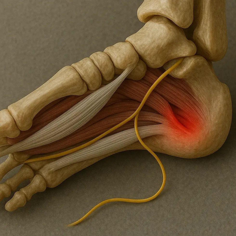
What Is Baxter’s Nerve?
Baxter’s nerve, also known as the first branch of the lateral plantar nerve, has both sensory and motor functions in the foot:
Motor function: Innervates the abductor digiti minimi muscle, which contributes to support of the foot arch.
Sensory function: Provides sensation to the region around the heel and the lateral part of the plantar fascia.
When this nerve becomes compressed or irritated, it can cause pain and reduced function in the heel region.
Why Is Baxter’s Nerve Entrapment Important?
The condition is often confused with plantar fasciitis because the symptoms may be similar. Patients with Baxter’s nerve entrapment often experience persistent pain that does not respond to standard plantar-fasciitis treatment. An accurate diagnosis is therefore essential for tailoring treatment and achieving effective pain relief.[2]
Clinically Relevant Anatomy
Baxter’s nerve, also known as the inferior calcaneal nerve, is the first branch of the lateral plantar nerve arising within the tarsal tunnel. The lateral plantar nerve contains both sensory and motor components:
Sensory components: Innervate the calcaneal periosteum, the long plantar ligament and the skin of the lateral plantar surface.
Motor components: Innervate the abductor digiti minimi, flexor digitorum brevis and quadratus plantae muscles.
The first branch of the lateral plantar nerve usually arises from the lateral plantar nerve near the bifurcation of the tibial nerve, but it may also arise from the tibial nerve before the bifurcation. The nerve follows a complex course:
Through the fascia: It passes through the superficial fascia at the superior border of the abductor hallucis, where the fascia is thicker laterally because of reinforcement from interfascicular ligaments.
Between muscles: The nerve runs distally between the lateral abductor fascia and the medial border of quadratus plantae.
Around the calcaneus: When it reaches the inferior border of the abductor hallucis, it turns laterally, passes approximately 5.5 mm anterior to the medial calcaneal tuberosity or spur, and continues between quadratus plantae and flexor digitorum brevis before reaching its motor target, the abductor digiti minimi.[2]
Etiology
Baxter’s nerve is particularly vulnerable to entrapment because of its anatomy and course. Two main compression sites have been identified:
Junction between quadratus plantae and abductor hallucis:
The nerve passes between the medial border of quadratus plantae and the thick lateral fascia of abductor hallucis. Tight fascial structures may cause compression.
Medial calcaneal tuberosity and/or heel spurs:
As the nerve passes anterior to the medial calcaneal tuberosity, increased contact pressure caused by heel spurs or muscular hypertrophy may contribute to entrapment.
Pronation of the rearfoot and midfoot may also create an unfavorable biomechanical position that increases pressure on the nerve as it turns laterally.[2]
This combination of anatomical and biomechanical factors makes Baxter’s nerve vulnerable to compression, which may cause chronic heel pain that is frequently mistaken for plantar fasciitis.
Clinical Presentation
Patients with Baxter’s nerve entrapment may experience the following symptoms:
Tenderness over the origin of the abductor hallucis: This may reproduce laterally radiating discomfort and/or paresthesia.[2]
Sharp, radiating pain: Pain typically worsens at night and after physical activity such as walking or running.[3]
Moderate muscle atrophy: Noticeable atrophy of the abductor digiti minimi may occur.[4]
Pain over the medial calcaneus: Pain is localized to the heel, particularly on the medial side.[4]
Examination
Biomechanical Assessment
During biomechanical assessment, the clinician commonly observes an overpronated foot posture. Pronation may contribute to nerve entrapment.[2]
Special Tests
Tinel’s test:
The test is performed by lightly tapping over the nerve at the suspected compression site. A positive test produces pain or tingling radiating along the course of the nerve.[3]
Phalen’s test:
The test involves passive inversion and plantar flexion of the foot, which may provoke pain by compressing the nerve.[4]
Windlass test:
Used to help rule out plantar fasciitis as a differential diagnosis.
Diagnostic Imaging
Radiography and bone scan:
Plain radiographs and bone scans may be used to exclude osseous pathology such as heel spurs or stress fractures.[2]
Serological Tests
Serological testing may be performed to exclude systemic arthropathy, such as rheumatoid arthritis or gout, as the cause of symptoms.[2]
This comprehensive clinical examination and use of special tests, together with imaging and serological testing, help confirm the diagnosis of Baxter’s nerve entrapment and exclude other potential causes of heel pain.
Differential Diagnoses for Baxter’s Nerve Entrapment
Several conditions can produce symptoms similar to Baxter’s nerve entrapment, and these should be considered when establishing an accurate diagnosis:
Plantar fasciitis:
A common cause of heel pain, usually more localized to the medial heel border and associated with weight-bearing.
Seronegative arthritis-related inflammation:
Rheumatic conditions such as psoriatic arthritis may cause inflammation in the heel region.
Tarsal tunnel syndrome:
Entrapment of the tibial nerve in the tarsal tunnel, producing radiating pain along the medial plantar surface of the foot.
Medial calcaneal neuritis:
Inflammation or irritation of the medial calcaneal branch of the tibial nerve.
Heel spurs:
May cause pain because of pressure or inflammation in tissues surrounding the spur.
Trauma:
For example, direct impact to the heel may injure a nerve or muscle.
Heel fat-pad atrophy:
Loss of cushioning in the heel fat pad may cause pain during weight-bearing.
Calcaneal stress fractures:
Stress fractures can cause pain that worsens with activity.
Periosteal inflammation:
Inflammation of the calcaneal periosteum may produce similar symptoms.
Treatment and Management of Baxter’s Nerve Entrapment
Medical Treatment
NSAIDs (nonsteroidal anti-inflammatory drugs):
May help reduce pain and inflammation.
Antiepileptic medications:
May be used to reduce neuropathic pain.[3]
Conservative Treatment
Kinesiology taping and orthoses:
Taping or custom orthoses may help reduce pressure on the nerve.
Stretching and strengthening exercises:
Focus on stretching the abductor hallucis and strengthening the intrinsic foot musculature.
Injection-Based Treatments:
Cryoablation and radiofrequency treatment: Reduce transmission of pain signals through the nerve.
Hydrodissection with 5% dextrose: Used to create space around the nerve.
Ultrasound-guided corticosteroid injection: May provide short-term relief of pain and inflammation.[3]
Surgical Treatment
Nerve decompression:
Performed when conservative treatment does not provide adequate symptom relief. The procedure releases the nerve from surrounding tissues causing compression.[1]
Early intervention and appropriate differential diagnosis are essential for effective management of Baxter’s nerve entrapment and prevention of chronic symptoms.
Sources:
Sahoo RK, Peng PW, Sharma SK. Ultrasound-Guided Hydrodissection for Baxter’s Neuropathy Secondary to Plantar Fasciitis: A Case Report. A&A Practice. 2020 Nov 1;14(13):e01339.
Stephen Offutt DP, Patrick DeHeer DP. How to address Baxter’s nerve entrapment. Podiatry Today. 2004 Nov 3;17(11).
Allam AE, Chang KV. Plantar Heel Pain. [Updated 2022 Sep 5]. In: StatPearls [Internet]. Treasure Island (FL): StatPearls Publishing; 2023 Jan-. Available from: https://www.ncbi.nlm.nih.gov/books/NBK499868/.
Prem A, Anand S. Baxter's nerve injury: an often overlooked cause of chronic heel pain: a case report. Arch Clin Exp Orthop. 2023;7:003-004.
Michigan Foot Doctors. BEST Heel Nerve Pain Treatment [Baxter’s Nerve Entrapment Treatment]. Available from: https://www.youtube.com/watch?v=1ZqubXExmc4 [last accessed 6/11/2021].


