

Elbow Osteoarthritis

Elbow osteoarthritis is a degenerative joint disorder that mainly affects older adults or people with a history of trauma to the elbow. Unlike large weight-bearing joints such as the hip and knee, osteoarthritis of the elbow is relatively uncommon. When it does occur, it is most often associated with long-term loading, repetitive use, or late complications following fractures or dislocations. The most characteristic symptoms are gradually increasing pain and reduced range of motion, which can eventually interfere with daily activities such as lifting, carrying, and rotational movements of the forearm¹,².
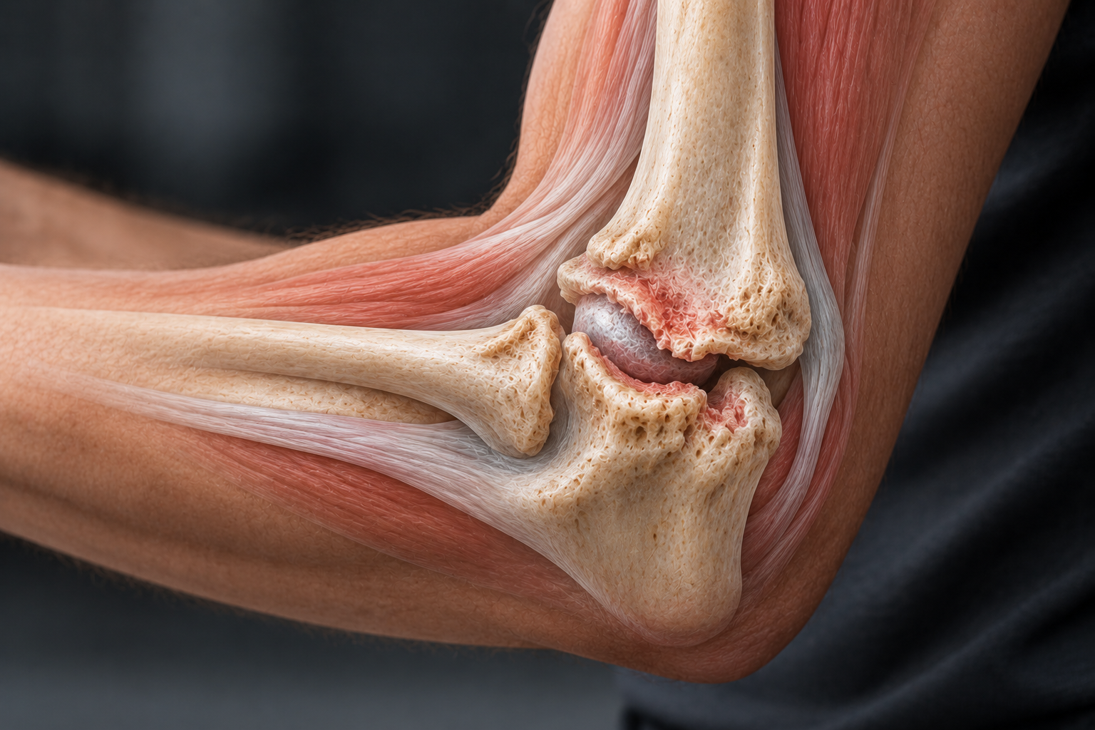
A key factor in the development of elbow osteoarthritis is altered biomechanics after injury. Even after successful fracture healing, joint-surface incongruity or ligamentous instability can create abnormal loading patterns. This may accelerate degenerative changes in the articular cartilage and surrounding structures, leading to osteophyte formation, synovial inflammation, and capsular thickening³,⁴,⁵.
Clinically Relevant Anatomy
The elbow is a true synovial hinge joint involving three bones: the distal humerus and the proximal ulna and radius. Anatomically, the joint consists of three separate but functionally connected components: the humeroulnar joint, the humeroradial joint, and the proximal radioulnar joint. The articular surfaces allow flexion and extension, as well as forearm rotation through interaction with the surrounding musculature.
The lateral deviation between the humerus and ulna, known as the carrying angle, is usually 10–15° in men and 20–25° in women⁶. This is important when assessing varus or valgus deformity associated with osteoarthritic changes.
Pathophysiology
Two main forms of elbow osteoarthritis are recognized: primary and post-traumatic⁷,⁸,⁹.
1. Primary Elbow Osteoarthritis
Primary osteoarthritis is rare and is seen almost exclusively in people exposed to long-term mechanical loading of the elbow, such as athletes, carpenters, bricklayers, and others with manual occupations. The condition is often unilateral and usually affects the dominant arm. Degenerative changes typically begin peripherally around the olecranon and coronoid processes, while the central articular surface is relatively preserved in the early stages. This initially causes pain at end-range flexion and extension, while mid-range positions may remain pain-free.
As the condition progresses, osteophyte formation, synovial thickening, and capsular contracture can reduce motion throughout the range and cause persistent pain.
2. Post-Traumatic Elbow Osteoarthritis
This is the most common form of elbow osteoarthritis. It develops after previous fractures, dislocations, or intra-articular injuries. Malunited fractures or chronic instability after ligament injury can alter load distribution across the joint surfaces and accelerate cartilage degeneration. Reduced joint congruity and possible micromotion may contribute to osteophyte formation, sclerosis, and subchondral cysts.
Patients with post-traumatic osteoarthritis often report pain and crepitus during both flexion-extension and rotational movements, and a history of previous injury is typical.
Epidemiology
Elbow osteoarthritis is much less common than osteoarthritis in larger joints. In a historical study by Ruelle et al. from 1965, only 0.56% of patients attending a rheumatology clinic had elbow osteoarthritis¹⁰. Many reported only mild symptoms, and two thirds had no pain. The most common clinical finding was reduced range of motion, particularly loss of extension early in the disease, while pronation and supination were often affected later.
Ulnar neuropathy was reported in approximately 15.5% of patients and may result from compression caused by osteophytes or thickened synovial tissue within the ulnar groove.
Clinical Presentation
The most prominent symptom of elbow osteoarthritis is pain that usually worsens with loading and movement, especially at the end ranges. In early stages, pain is typically provoked by full flexion or extension, whereas in more advanced disease it may occur throughout the entire range of motion. Pain is often accompanied by stiffness, reduced mobility, and progressive functional impairment of the arm and hand.
In post-traumatic osteoarthritis, loss of extension is the most common finding because of previous injury followed by osteophyte formation or capsular contracture. In primary osteoarthritis, the pattern of motion loss is more variable. A thorough history is therefore important to identify previous trauma, surgery, and loading history.
Ulnar neuropathy is a common complication of elbow osteoarthritis and may result from nerve compression in the ulnar groove, particularly in the presence of osteophytes or capsular thickening. Clinical assessment should include symptoms such as reduced sensation in the ring and little fingers, weakness of the intrinsic hand muscles, altered grip strength, and neuropathic pain. Red flags include suspicion of septic arthritis, particularly in patients with previous surgery, joint prostheses, or immunosuppression.
Diagnostic Assessment
Clinical examination should begin with inspection and comparison of the affected arm with the contralateral side. Assessment should include swelling, muscle atrophy, malalignment, and visible deformity. Palpation is used to identify joint-line tenderness and thickening. Active and passive range-of-motion testing assesses motion loss and pain at specific joint angles. The shoulder and wrist should also be examined to exclude referred pain.
Neurovascular examination is essential, with particular attention to ulnar nerve function. Sensation in the ulnar distribution should be tested, together with strength of the dorsal interossei and adductor pollicis.
Ligament stability is assessed with varus and valgus stress testing. Laxity may indicate concurrent ligament injury or previous trauma that predisposes to unstable osteoarthritis.
Crepitus is commonly present during movement, especially in advanced osteoarthritis.
Diagnostic Imaging:
X-ray is the first-line imaging method. Standard views usually include anteroposterior, lateral, and oblique projections. Early findings include anterior and posterior osteophytes, often with preservation of the central joint space. In patients with previous injury, imaging should assess malalignment, radiocapitellar osteoarthritis, and possible heterotopic ossification.
CT may be used when complex pathology is suspected, such as intra-articular loose bodies, malunited fractures, or heterotopic ossification.
MRI has a limited role in routine elbow osteoarthritis but may be useful when the differential diagnosis is unclear.
Outcome Measures
The following scales may be used to assess function and treatment outcomes:
American Shoulder and Elbow Surgeons (ASES) Elbow Outcome Score
Broberg and Morrey Elbow Scale
Mayo Clinic Performance Index for the Elbow
Hospital for Special Surgery (HSS) Assessment Scale
Liverpool Elbow Score¹¹
Treatment and Management
Conservative Treatment is the first-line approach for both primary and post-traumatic elbow osteoarthritis. It may include:
Medication, including NSAIDs and intra-articular corticosteroid injections.
Braces and elastic supports may provide symptom relief and improve proprioception.
Physiotherapy, although specific literature for elbow osteoarthritis is limited, generally follows the same principles used for other joints: range-of-motion exercises, strengthening, patient education, and activity modification. Manual therapy may also be useful.
If conservative treatment does not provide adequate relief, surgery may be considered.
Surgical Options:
Arthroscopic debridement to remove osteophytes and loose bodies³
Ulnohumeral decompression or interposition arthroplasty
Total elbow arthroplasty (TEA) – including:
Linked or semi-constrained prosthesis (stable)
Unlinked or resurfacing prosthesis (requires intact ligaments)
Convertible prosthesis (can be converted to a linked design if needed)
Radial head resection or radiocapitellar arthroplasty
Arthrodesis is a last-resort option for severe, disabling osteoarthritis in patients with low functional demands¹²
Differential Diagnoses
For elbow pain and reduced function, the following conditions should be considered and excluded when appropriate:
Ligament injuries (MCL/LCL)
Epicondylar tendinopathies (lateral/medial)
Ulnar neuropathy
Occult fracture
Referred pain from the cervical spine
Rehabilitation
The goals of rehabilitation are to reduce pain and inflammation, improve or maintain mobility, and strengthen the surrounding musculature.
Non-Surgical Management:
Thermotherapy for pain relief
Range-of-motion exercises and stretching to maintain mobility
Gradual strengthening of the elbow, shoulder, and upper back
Progression to a structured home-exercise program over 4–6 weeks
After Surgery:
Initial focus on pain relief and edema reduction
The elbow is often bandaged or immobilized during the early phase
Gradual progression of passive and active range of motion
Progressive strengthening to restore function
Full rehabilitation may take up to 3 months and often requires close follow-up
Sources:
Athwal G, Fischer S. Osteoarthritis of the elbow - orthoinfo - aaos. 2017: https://orthoinfo.aaos.org/en/diseases--conditions/osteoarthritis-of-the-elbow/
Versus Arthritis. Osteoarthritis (OA) of the elbow and shoulder. https://www.versusarthritis.org/about-arthritis/conditions/osteoarthritis-oa-of-the-elbow-and-shoulder/
Cheung EV, Adams R, Morrey BF. Primary osteoarthritis of the elbow: current treatment options. JAAOS-Journal of the American Academy of Orthopaedic Surgeons. 2008 Feb 1;16(2):77-87.
Gramstad GD, Galatz LM. Management of elbow osteoarthritis. JBJS. 2006 Feb 1;88(2):421-30.
Doherty MI, Preston BR. Primary osteoarthritis of the elbow. Annals of the rheumatic diseases. 1989 Sep 1;48(9):743-7.
Palastanga NP, Soames RW, Anatomy and human movement: Structure and function. 6th ed. Edinburgh: Churchill Livingstone; 2011 Sep 27. ISBN: 9780702035531.
2. Biswas D, Wysocki RW, Cohen MS. Primary and Posttraumatic arthritis of the elbow. Arthritis. 2013;2013:1–6.
Cheung EV, Adams R, Morrey BF. Primary osteoarthritis of the elbow: current treatment options. JAAOS-Journal of the American Academy of Orthopaedic Surgeons. 2008 Feb 1;16(2):77-87.
Ravalli S, Pulici C, Binetti S, Aglieco A, Vecchio M, Musumeci G. An overview of the pathogenesis and treatment of elbow osteoarthritis. Journal of Functional Morphology and Kinesiology. 2019 Jun;4(2):30.
Ruelle JL, Debeuckelaere M, Raynal L. Introduction à l’étude ducoude. Journal belge de rhumatologie et de médecine physique 1965;20:161–7
Smith MV, Calfee RP, Baumgarten KM, Brophy RH, Wright RW. Upper Extremity-Specific Measures of Disability and Outcomes in Orthopaedic Surgery. The Journal of Bone and Joint Surgery American volume. 2012;94(3):277-285. doi:10.2106/JBJS.J.01744.
Orthogate. Osteoarthritis of the Elbow. September 4, 2015: https://www.orthogate.org/patient-education/elbow/osteoarthritis-of-the-elbow


