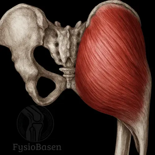
Gluteus Medius
- Fysiobasen

- Jan 11
- 9 min read
The gluteus medius is a large, fan-shaped muscle located on the lateral aspect of the hip and the upper part of the gluteal region. It plays a central role in pelvic stabilization during walking and running and functions as the primary hip abductor.

Origin
Gluteus medius has a broad origin from the lateral surface of the ilium. This area lies between the anterior and posterior gluteal lines, which serve as clear anatomical landmarks on the hip bone.The origin extends widely from the iliac crest (crista iliaca) inferiorly toward the greater sciatic notch (incisura ischiadica major) and also includes the gluteal aponeurosis, a thick connective tissue layer linking the gluteal muscles with each other and adjacent structures.
Insertion
From its broad origin, the muscle fibers of gluteus medius run in three distinct directions before converging into a common, thick, flat tendon:
Posterior fibers run obliquely forward and downward
Middle fibers run vertically downward
Anterior fibers run obliquely backward and downward
All fibers unite into a strong, flat tendon that inserts on the lateral surface of the greater trochanter of the femur. The insertion lies on a rough, well-defined superolateral facet of the greater trochanter.
Between the tendon and the greater trochanter lies the trochanteric bursa, which reduces friction and allows smooth tendon movement over bone.
Location and Anatomical Relations
Gluteus medius lies deep to gluteus maximus, the largest muscle of the gluteal region. However, gluteus maximus does not fully cover gluteus medius; approximately one third of its anterosuperior portion lies directly beneath the skin.
This area, known as the upper outer quadrant of the gluteal region, is commonly recommended for intramuscular injections, as it carries a low risk of injury to major nerves and vessels.
Medially and deep to gluteus medius lies gluteus minimus, while laterally the muscle borders the iliotibial tract. Posteriorly, it is adjacent to piriformis, another key hip stabilizer.Between gluteus medius and minimus pass the superior gluteal nerve and artery.
Shape and Fiber Orientation
Gluteus medius is classically described as fan-shaped, being widest at its origin and narrowing distally toward its tendon. The variation in fiber orientation allows the muscle to perform different actions depending on which portions are preferentially activated.
Deeper Anatomical Understanding (Topography and Palpation)
On palpation, the muscle can be easily identified by locating the iliac crest and moving the fingers inferiorly along the lateral hip. During active hip abduction, such as lifting the leg sideways, the contraction of gluteus medius is clearly palpable.
On ultrasound and MRI, gluteus medius appears as a distinct structure between gluteus maximus and minimus, with a clear interface at the greater trochanter. Thickening or inflammation of the underlying bursa may be visible and suggests irritation or injury.
Function
Gluteus medius is primarily responsible for hip abduction, moving the leg away from the body’s midline. In addition, different portions of the muscle contribute to specific movements:
Anterior fibers: Assist in hip flexion and internal rotation
Posterior fibers: Assist in hip extension and external rotation, particularly at small hip angles
Entire muscle: Hip abduction and, most importantly, lateral pelvic stabilization during gait and running
When one foot is lifted during walking, gluteus medius on the stance leg contracts strongly to prevent the pelvis from dropping on the opposite side. This function is essential for normal gait, running, and all single-leg balance activities.
Gluteus medius also acts as an important antagonist to the hip adductors and is crucial for maintaining a neutral pelvic position in daily activities, sports, and functional training.
Clinical Importance and Context
Weakness or inhibition of gluteus medius is associated with several orthopedic conditions and movement dysfunctions, including:
Trendelenburg gait
Iliotibial band syndrome
Anterior knee pain (patellofemoral pain syndrome)
Low back pain related to pelvic instability
Routine assessment of strength, control, and activation of this muscle is therefore essential in clinical practice, especially in athletes, individuals with hip pain, and patients recovering from hip or knee injuries.
Innervation
Gluteus medius is innervated by the superior gluteal nerve, arising from the sacral plexus, with root values L4, L5, and S1.
The superior gluteal nerve exits the pelvis through the suprapiriform foramen, just above the piriformis muscle, before dividing into branches that innervate gluteus medius, gluteus minimus, and tensor fasciae latae.
Injury to this nerve leads to significant pelvic instability and gait disturbance. The classic clinical sign is a positive Trendelenburg sign, where the pelvis drops on the contralateral side during single-leg stance.
Blood Supply
Gluteus medius receives its primary blood supply from the superior gluteal artery, a branch of the internal iliac artery. This artery divides into:
A deep branch, supplying the muscle belly
A superficial branch, supplying surrounding connective tissues
The tendon of gluteus medius is supplied by the trochanteric anastomosis, formed by branches of the medial circumflex femoral artery, superior gluteal artery, and inferior gluteal artery.This robust vascular network is important for healing following overuse injuries or surgery.
Detailed Functional Role
Gluteus medius performs multiple, partially opposing actions due to its segmented fiber architecture:
Hip Abduction
The primary action of the entire muscle, essential in both isolated movements and complex activities such as walking, running, and jumping.
Hip Rotation
Anterior fibers: Internal rotation and assistance in hip flexion
Posterior fibers: External rotation and assistance in hip extension
At small hip flexion angles (0–25°), external rotation predominates. At 90° of hip flexion, the muscle’s capacity for internal rotation increases dramatically, becoming up to eight times stronger than at lower flexion angles.
Pelvic Stabilization
Clinically the most important function. During single-leg stance, gluteus medius stabilizes the pelvis in the frontal plane. Weakness results in pelvic drop and the characteristic Trendelenburg gait.
Synergy With Other Muscles
Gluteus medius functions as part of a coordinated muscular system:
Gluteus minimus and tensor fasciae latae: Assist in abduction, internal rotation, and lateral pelvic stability
Contralateral quadratus lumborum: Forms a lateral stabilization sling; weakness in gluteus medius often leads to overactivity and low back pain
Gluteus maximus: Works with posterior fibers during hip extension and external rotation
Balanced interaction among these muscles is essential for optimal hip mechanics and injury prevention.
Mobilizing vs. Stabilizing Function
Gluteus medius uniquely combines stabilizing and mobilizing roles.During single-leg, weight-bearing tasks it functions primarily as a stabilizer, while during running, jumping, and directional changes it contributes to both force production and movement control.
Role in Daily Activities and Load-Bearing Tasks
Gluteus medius is essential for:
Walking and running: Prevents lateral pelvic drop
Stair ascent and descent: Controls hip stability under load
Lateral movements: Critical in sports with rapid direction changes
Single-leg balance tasks: Key for injury prevention and functional control
Clinical Findings and Typical Symptoms of Dysfunction
Dysfunction of gluteus medius may result from weakness, inhibition, overuse, or injury. Common findings include:
Lateral hip pain, often over the greater trochanter
Reduced strength and endurance during single-leg tasks
Positive Trendelenburg sign
Altered gait, often with compensatory trunk lean (Duchenne gait)
Myofascial trigger points with referred pain along the lateral thigh
Palpation, Examination, and Pain
During clinical examination, palpation and specific functional tests are essential:
Palpation
The muscle is best palpated by placing the hand laterally just below the iliac crest, directly above the greater trochanter.In a side-lying position with active hip abduction, the muscle contraction can be clearly felt.Tenderness on palpation over the insertion at the greater trochanter may indicate gluteus medius tendinopathy or trochanteric bursitis.
Muscle Strength
Strength is best assessed using side-lying hip abduction, as well as single-leg stance and mini-squats, while observing pelvic stability and control.
Trendelenburg Test
The patient stands on one leg for 30 seconds while the clinician observes for pelvic drop on the contralateral side, indicating insufficient lateral pelvic stabilization.
Relevance for Sport, Rehabilitation, and Physiotherapy
Gluteus medius plays a key role in sport performance, rehabilitation, and injury prevention:
Prevention of Knee Injuries
Weakness of gluteus medius is associated with an increased risk of ACL injuries and patellofemoral pain syndromes.Improved activation and strengthening of gluteus medius significantly reduces injury rates in the lower extremity.
Running-Related Injuries
Conditions such as iliotibial band syndrome (ITBS), trochanteric bursitis, and hip-related pain in runners are strongly correlated with gluteus medius weakness or dysfunction.
Rehabilitation After Surgery or Injury
Following hip and knee surgeries, including hip arthroscopy and knee procedures (e.g., ACL reconstruction), targeted gluteus medius strengthening is a core component of rehabilitation programs.
Sports Performance
Athletes in football, handball, basketball, and athletics rely heavily on a strong and well-activated gluteus medius for force transfer, balance, and performance, particularly during rapid direction changes and explosive movements.
Postural Function
Gluteus medius has a critical postural role:
It stabilizes the pelvis in the frontal plane and prevents pelvic drop during single-leg loading, which is essential during both static postures (e.g., prolonged standing) and dynamic movements (e.g., walking, running, jumping).
With weakness or inhibition, compensatory overactivity often develops in the tensor fasciae latae, quadratus lumborum, and hip adductors. This may lead to muscle imbalances, chronic pain, and reduced postural control.
Over time, such compensation patterns can contribute to structural adaptations, including functional scoliosis, increased lumbar lordosis, or asymmetrical lower-limb loading.
Relation to Movement Patterns, Faulty Loading, and Compensation
Weak or inhibited gluteus medius leads to immediate and long-term compensatory movement strategies:
Increased hip adduction and internal rotation during activity, increasing stress on structures such as the iliotibial band, menisci, and patellofemoral joint.
Increased spinal and pelvic loading, as the body compensates via quadratus lumborum and spinal extensors to stabilize the pelvis.
Lateral shift of the center of mass, causing abnormal loading of the knees, ankles, and foot arches, increasing the risk of plantar fasciitis, medial tibial stress syndrome, and chronic knee pain.
Increased injury risk during landings and direction changes, particularly in sports, due to reduced stability and impaired neuromuscular control.
Exercises for Gluteus Medius
Gluteus medius is a key muscle for pelvic stability, efficient gait and running mechanics, and injury prevention in the lower extremity.The exercises below are research-based and demonstrate high muscle activation in EMG studies.
Side Plank With Hip Abduction (“Side Bridge”)
Primary activation: Gluteus medius
Secondary activation: Quadratus lumborum, obliques, gluteus minimus, tensor fasciae latae
Execution:
Lie on your side with the elbow directly under the shoulder
Body aligned in a straight line, legs stacked
Lift the body so weight is supported on the forearm and lateral foot
Slowly raise the top leg, hold briefly, then lower with control
Dosage: 10–15 repetitions per side, 2–3 sets
Effect: EMG studies show up to 74% MVIC activation of gluteus medius, making this one of the most effective isolated exercises for this muscle. Best suited for higher functional levels or late-stage rehabilitation.
Clinical use: Ideal for athletes and active individuals. Should be introduced gradually after hip, knee, or ankle injury or surgery.
Variations:
Easier: Knees bent (shorter lever arm)
Advanced: Add resistance band around thighs
Single-Leg Squat
Primary activation: Gluteus medius
Secondary activation: Quadriceps, hamstrings, gluteus maximus, core muscles
Execution:
Stand on one leg, opposite leg lifted forward
Maintain neutral spine and upright trunk
Slowly bend hip and knee to ~45° while keeping pelvis level
Push back up using hip and gluteal musculature
Dosage: 10–12 repetitions per side, 2–3 sets
Effect: Approximately 64% MVIC activation of gluteus medius, providing excellent functional transfer to sport and daily activity.
Clinical use: Highly effective for functional rehabilitation and injury prevention, especially after ACL reconstruction, knee instability, and hip disorders.
Variations:
Easier: Light support from wall or chair
Advanced: Unstable surface or added external load
Side-Lying Hip Abduction
Primary activation: Gluteus medius
Secondary activation: Gluteus minimus, tensor fasciae latae
Execution:
Lie on your side with hips stacked and body aligned
Lift the top leg upward without rotating the pelvis
Lower the leg slowly with control
Dosage: 12–15 repetitions per side, 2–3 sets
Effect: EMG activity around 56% MVIC. Safe and effective early in rehabilitation and suitable for most functional levels.
Clinical use: Often used as a first-line exercise for gluteus medius weakness, especially in older adults, post-surgical patients, or after prolonged immobilization.
Variations:
Easier: Slight hip and knee flexion
Advanced: Resistance band or ankle weight
Single-Leg Bridge
Primary activation: Gluteus medius and gluteus maximus
Secondary activation: Hamstrings, core muscles
Execution:
Lie on your back with knees bent and feet on the floor
Extend one leg forward
Lift the pelvis by pressing the heel of the stance leg into the floor
Keep pelvis level and stable
Lower slowly with control
Dosage: 10–15 repetitions per side, 2–3 sets
Effect: EMG studies show approximately 47% MVIC activation of gluteus medius. Effective for strengthening both gluteus medius and maximus and improving pelvic stability.
Clinical use: Well suited for rehabilitation following hip or low-back injury and for general pelvic stability training.
Variations:
Easier: Both feet on the floor
Advanced: Add resistance band around knees
Lateral Step-Up
Primary activation: Gluteus mediusSecondary activation: Quadriceps, gluteus maximus, hamstrings
Execution:
Stand sideways next to a step or low box
Place the nearest foot on the step
Press through the foot to lift the body while keeping the pelvis level
Lower slowly with control
Dosage: 12–15 repetitions per side, 2–3 sets
Effect: Produces approximately 41% MVIC activation and has strong functional transfer to walking, stair climbing, and lateral sports movements.
Clinical use: Appropriate for knee rehabilitation (especially ACL), hip pathology, and general injury prevention.
Variations:
Easier: Lower step height
Advanced: Hold a dumbbell or kettlebell in the opposite hand
References
Moore, K. L., Dalley, A. F., & Agur, A. M. R. (2014). Clinically Oriented Anatomy (7th ed.). Philadelphia, PA: Lippincott Williams & Wilkins.
Palastanga, N., & Soames, R. (2012). Anatomy and Human Movement: Structure and Function (6th ed.). Edinburgh: Churchill Livingstone.
Standring, S. (2016). Gray’s Anatomy (41st ed.). Edinburgh: Elsevier Churchill Livingstone.