Serratus Anterior
- Fysiobasen

- Jan 11
- 5 min read
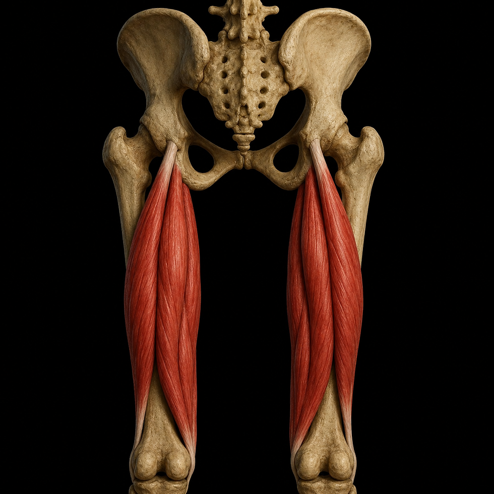
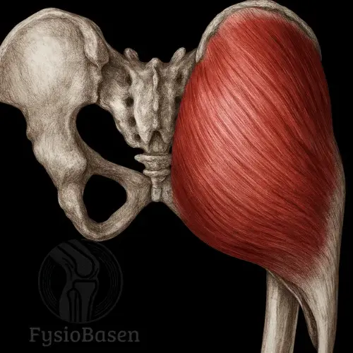
The serratus anterior is a fan-shaped, deep muscle that covers the lateral aspect of the thoracic cage and lies closely beneath the scapula. It has a characteristic “saw-toothed” appearance and is visible in well-trained individuals along the ribs beneath the axilla. The muscle is considered essential for scapular function, and weakness of the serratus anterior results in immediate functional impairment of the entire shoulder complex.

Origin
The serratus anterior is divided into three functional portions based on rib level and scapular attachment.
• Upper portion: from the 1st–2nd ribs, inserting at the superior angle of the scapula
• Middle portion: from the 2nd–3rd ribs, inserting along the medial border of the scapula
• Lower portion: from the 4th–9th ribs, occasionally the 10th, inserting at the inferior angle and medial border
The muscle fibers are organized into nine separate digitations. The lowest four interdigitate with the external oblique muscle of the abdomen. The fibers run obliquely posteriorly and superiorly toward the medial surface of the scapula, forming a multipennate pattern. The lower portion is the most powerful.
Insertion
The common insertion is the anteromedial surface of the scapula, including:
• The superior angle
• The medial border
• The inferior angle
By attaching to the anterior surface of the scapula, the serratus anterior holds the scapula firmly against the thoracic wall. This enables stable movement and rotation of the scapula during arm use above shoulder height.
Innervation
Nerve: Long thoracic nerve
Spinal roots: C5–C7
Mnemonic: “SALT = Serratus Anterior – Long Thoracic”
Clinical memory aid: “C5, 6, 7 – raise your arms to heaven”
The nerve runs superficially along the muscle, making it vulnerable to injury during surgical procedures, such as axillary lymph node dissection, or due to mechanical stress, for example carrying heavy loads or trauma to the thoracic wall.
Blood supply
The muscle receives a rich blood supply from several sources.
• Lateral thoracic artery as the primary source
• Superior thoracic artery
• Thoracodorsal artery, a branch of the subscapular artery
These arteries ensure adequate perfusion even during large scapular movements and when the muscle functions as an accessory inspiratory muscle during increased respiratory demand.
Structural relations
• Superficial: pectoralis major and pectoralis minor
• Posterior: the scapula, including the entire medial and inferior border
• Lateral and anterior: ribs 1–9
• Inferior: external oblique muscle
On palpation, the muscle is best accessed between the pectoralis major and latissimus dorsi, just posterior and slightly inferior to the axilla.
Function
Primary functions
The serratus anterior has three main roles in scapulothoracic mechanics.
• Protraction of the scapulaDraws the scapula forward and around the thoracic wall, for example during punching movements
• Upward rotation of the scapulaPulls the inferior angle laterally and superiorlyEssential for arm elevation above 90 degrees, in cooperation with the trapezius
• Stabilization of the scapula against the thoraxPrevents scapular wingingMaintains contact with the rib cage during dynamic movement
Secondary functions
• Scapular suspensionKeeps the scapula “floating” and correctly oriented for optimal glenohumeral articulation
• Antagonist to retractionBalances the rhomboids and middle trapezius during dynamic movements
• Accessory muscle of inspirationActivated when the scapula is fixed, for example during respiratory distress, asthma, or physical exertionElevates the ribs laterally and increases thoracic expansion
Synergistic relationships
Interaction with the trapezius (force couple)
The serratus anterior works closely with the upper and lower fibers of the trapezius to produce effective upward rotation of the scapula. In particular, the lower serratus anterior and lower trapezius act together to guide the inferior angle of the scapula laterally and upward. This force couple is essential for full overhead shoulder function and efficient arm elevation.
Interaction with pectoralis major, pectoralis minor, and latissimus dorsi
During pulling movements, rotational tasks, and fixation of the shoulder girdle, the serratus anterior cooperates with the pectoralis muscles and latissimus dorsi. In these contexts, the muscle contributes primarily to scapular positioning and stability rather than gross force production.
Clinical significance
The serratus anterior is essential for normal shoulder mechanics. Dysfunction or underactivity leads rapidly to loss of scapular control, reduced range of motion above 90 degrees of arm elevation, and a markedly increased risk of subacromial impingement and secondary rotator cuff pathology. Clinically, this often presents as weakened pushing movements, including bench press, push-ups, and throwing activities.
Common clinical problems
Scapula alata (medial winging)
Medial scapular winging most commonly results from injury to the long thoracic nerve. This may occur after axillary surgery, trauma to the lateral thoracic wall, or prolonged load from heavy backpacks. Patients typically demonstrate visible scapular protrusion during wall push tasks and have difficulty elevating the arm.
Serratus anterior myopathy and overuse
Overuse-related dysfunction is frequently observed in swimmers, boxers, and tennis players. Symptoms are often diffuse and poorly localized, presenting as pain along the lateral rib cage combined with reduced scapular control during dynamic arm movements.
Shoulder dysfunction due to underactivity
Insufficient serratus anterior activation leads to reduced upward rotation of the scapula, increasing the risk of subacromial impingement. Over time, compensatory overuse of the levator scapulae and upper trapezius commonly develops.
Function-based observation
During a push-up plus, insufficient scapular protraction indicates serratus anterior weakness. Symptom reduction during the scapular assistance test suggests underactivity of the muscle, while visible winging during a wall push test is a clear indicator of serratus anterior dysfunction.
Clinical testing
In the punch-out test, the patient presses the hand against a wall while the examiner observes scapular behavior. Medial border winging indicates serratus anterior insufficiency. During resisted protraction at 90–100 degrees of shoulder flexion with the elbow extended, weakness is evident if the scapula is displaced posteriorly. Similarly, resisted shoulder abduction in the scapular plane may reveal collapse of the scapula when serratus anterior function is inadequate.
Principles for training the serratus anterior
Training should emphasize proximal control, scapular stability, and functional integration with the rest of the shoulder girdle. Exercises must promote scapular protraction and upward rotation, reinforce stable contact between the scapula and thoracic wall to prevent winging, and preferentially target the lower fibers, which are most prone to underactivity.
EMG-based activation hierarchy
EMG studies demonstrate that the highest serratus anterior activation occurs during push-up plus, dynamic hug, and wall slide variations, with progressively lower activation during standing serratus punches and overhead shrug movements. These findings support the use of closed-chain and scapula-focused exercises in both rehabilitation and performance training.
Training progression
Phase 1 – Activation and isolation
In cases of pain, scapular winging, or early postoperative rehabilitation, training focuses on low-load activation and proprioceptive re-education. Wall press variations, supine shoulder protraction, and isometric protraction exercises are commonly used to re-establish neuromuscular control.
Phase 2 – Motor control and progression
As control improves, exercises such as modified push-up plus, dynamic hug with resistance, and wall slides with external rotation are introduced. These movements challenge scapular stability while maintaining high serratus anterior activation.
Phase 3 – Functional strength and eccentric control
In later stages, standing serratus punches, overhead shrug variations, bear crawls, and plank-based exercises are used to improve functional strength, eccentric control, and transfer to sport- or work-specific demands.
Load management and recovery
In cases of overactivity or pain, recovery strategies may include breathing-focused relaxation in side-lying to improve rib mobility, light dynamic scapular movements with resistance bands, and gentle serratus anterior foam rolling between the scapula and thorax while avoiding direct pressure on the ribs.
Summary
The serratus anterior is essential for scapular control, arm elevation, and rotational stability. Underactivity leads to scapular winging, impingement, and reduced upper-limb strength. EMG evidence consistently demonstrates highest activation during push-up plus, dynamic hug, and wall slide exercises. Effective training should integrate motor control, strength development, and functional transfer, with continuous attention to scapular movement quality.
Sources
Schünke M, Schulte E, Schumacher U. Prometheus: General Anatomy and Musculoskeletal System. 2nd ed. Thieme Verlag; 2007:294–295.
Muscolino JE. The Muscular System Manual: The Skeletal Muscles of the Human Body. 2nd ed. Elsevier Mosby; 2005:214–217.
Berlit P. Clinical Neurology. 3rd ed. Springer Verlag; 2011:345.
Standring S. Gray’s Anatomy. 41st ed. Elsevier Churchill Livingstone; 2016.