Gluteus Minimus
- Fysiobasen

- Jan 11
- 8 min read
The gluteus minimus is the smallest and deepest of the three gluteal muscles, located on the posterolateral aspect of the hip region. Together with the gluteus medius and gluteus maximus, it forms the lateral musculature of the buttock and is part of the functional complex responsible for movement and stability of the hip joint. Gluteus minimus plays a particularly important role in pelvic control during gait and single-leg stance and therefore has high clinical relevance in both rehabilitation and injury prevention.

Origin and Insertion
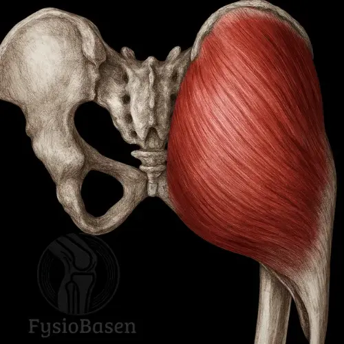
The gluteus minimus originates from the lateral (external) surface of the ilium, specifically between the anterior gluteal line (linea glutea anterior) and the inferior gluteal line (linea glutea inferior). The muscle fibers form a triangular pattern that converges into a narrow tendon.
This tendon runs in an anteroinferior direction and passes over the superior surface of the greater trochanter of the femur. Here, it is separated from the bone by a small bursa—the trochanteric bursa of the gluteus minimus—and ultimately inserts on the anterolateral aspect of the greater trochanter. In some anatomical variants, muscle fibers may blend with adjacent structures such as the piriformis, the superior gemellus muscle, or even the vastus lateralis.
Topographical Relations
The gluteus minimus lies directly against the inner surface of the hip bone (ilium) and is therefore in contact with bone across its entire deep surface. It is covered externally by the gluteus medius. Between these two muscles run both the superior gluteal artery and the superior gluteal nerve, an important consideration during surgical procedures in the hip region.
Anteriorly, the gluteus minimus borders the tensor fasciae latae muscle, while posteriorly it relates to the piriformis muscle. At its insertion on the greater trochanter, the tendon of the gluteus minimus lies over the reflected tendon of the rectus femoris and the hip joint capsule—an anatomical detail that may be relevant in intra-articular hip conditions.
Innervation
The gluteus minimus is innervated by the superior gluteal nerve, a motor nerve originating from the sacral plexus (nerve roots L4–S1). The nerve travels together with the superior gluteal artery through the suprapiriform space of the greater sciatic foramen and then branches between the gluteus medius and gluteus minimus as it courses anteriorly.
The innervation is consistent and specific, making the superior gluteal nerve clinically important in both neurological assessment and surgical interventions in the pelvic and gluteal regions. Lesions or compression of this nerve can lead to isolated weakness of the gluteus minimus and medius, with clear consequences for gait.
Blood Supply
The gluteus minimus receives its arterial supply primarily from the superior gluteal artery, a branch of the internal iliac artery. The deep branch of this artery runs between the gluteus medius and minimus and supplies both muscles.
In addition, the trochanteric anastomosis contributes to the blood supply of the distal portions of the muscle and its tendon insertion. This anastomosis includes branches from:
the superior gluteal artery
the inferior gluteal artery
the medial circumflex femoral artery
A robust blood supply to the tendon insertion is particularly important in cases of tendon injury or surgical repair in this region.
Function
The gluteus minimus has several functions that vary depending on the position of the hip joint and which portions of the muscle are activated:
1. Hip Abduction
When the pelvis is stabilised and the muscle’s origin is fixed, the gluteus minimus draws the femur laterally, away from the body’s midline. This contributes to lateral movement of the leg, which is an essential component of balance and gait.
2. Internal Rotation of the Hip
When the anterior muscle fibers are selectively activated, particularly when the hip joint is flexed, the movement shifts from abduction to rotation. This is due to a change in the vector and angle of the muscle’s line of pull and results in internal (medial) rotation of the femur.
3. Pelvic Stabilisation During Gait
Perhaps the most clinically relevant function is that the gluteus minimus, together with the gluteus medius, prevents the pelvis from dropping toward the contralateral side during the stance phase of gait. When standing on one leg, the ipsilateral abductors (minimus and medius) must pull the ilium down toward the greater trochanter to keep the pelvis level.
If the muscle is weakened or inactive, the pelvis will drop on the side opposite the stance leg—a sign known as the Trendelenburg sign.
Clinical Significance of the Gluteus Minimus
The primary clinical role of the gluteus minimus is stabilisation of the pelvis during single-leg weight bearing, a function that is essential for normal walking and running. Failure of this stabilising mechanism results in a characteristic gait pattern known as Trendelenburg gait, in which the pelvis drops toward the contralateral side of the stance leg.
The Trendelenburg sign is not solely an expression of gluteus medius weakness, as is often assumed, but also reflects dysfunction of the gluteus minimus. This is supported by electromyographic studies demonstrating significant minimus activity during single-leg loading and the swing phase of gait.
In lumbopelvic dysfunctions, such as instability of the sacroiliac joint or rotational disturbances of the pelvic ring, the gluteus minimus may also compensate for weaknesses elsewhere. This compensatory role can lead to overactivity or overload of the muscle.
Common Injuries and Conditions
1. Gluteus Minimus Tendinopathy and Greater Trochanteric Pain Syndrome (GTPS)
A common but often overlooked condition is tendinopathy of the gluteus minimus. This can cause pain over the lateral hip and is easily mistaken for trochanteric bursitis or referred lumbar pain. Together with the gluteus medius, the minimus is the most frequently affected muscle in GTPS.
GTPS primarily affects women over the age of 40 and is often associated with pre-existing hip osteoarthritis, lumbar spine disorders, or biomechanical deviations such as increased hip adduction during the stance phase of gait. MRI typically reveals degenerative changes in the tendon, fluid accumulation around the greater trochanter, and, in some cases, partial tendon tears.
2. Trigger Points and Referred Pain
The gluteus minimus contains several distinct trigger points, particularly in the anterior and middle fiber segments. These can produce referred pain extending down to the lateral calf and foot, which may be mistaken for sciatica. The key differentiating features are the absence of neurological deficits and a negative straight leg raise (Lasègue) test.
With chronic overload—for example from running on cambered surfaces, unilateral weight bearing, or asymmetric gait—these trigger points may remain persistently active and result in significant functional impairment.
3. Postoperative Atrophy and Reduced Function
Following hip arthroplasty and in degenerative conditions such as hip osteoarthritis (coxarthrosis), atrophy of the gluteus minimus is frequently observed, particularly in the anterior portion of the muscle. This segment is critical for internal rotation and control of the hip under load. Studies have shown that minimus atrophy is associated with reduced balance, increased fall risk, and altered muscle recruitment during functional movements.
Biomechanical Context and Interaction With Other Muscles
The gluteus minimus never functions in isolation. It works synergistically with the gluteus medius to draw the greater trochanter toward the iliac crest, thereby stabilising the pelvis.
During gait, it also cooperates with:
Tensor fasciae latae to control hip adduction and internal rotation of the femur
Quadratus lumborum to counteract pelvic drop in the frontal plane
Iliacus and psoas major during the transition from stance to swing phase to initiate hip flexion
If the gluteus minimus is weak, these synergistic relationships are disrupted, and compensatory movement patterns may lead to secondary symptoms in the lower back, knee, or ankle.
In strength training, the gluteus minimus should be trained separately from the gluteus maximus. The maximus primarily functions as a hip extensor and external rotator, whereas the minimus acts as an abductor and internal rotator. This distinction is crucial for targeted rehabilitation, particularly following surgery or in cases of pelvic pain.
Strength Exercises for the Gluteus Minimus: Evidence-Based Activation and Rehabilitation
The gluteus minimus plays a decisive role in hip joint stability, especially during single-leg activities such as walking, stair climbing, and running. It is often neglected in training and rehabilitation, yet recent EMG and MRI studies have shown that dysfunction of this muscle contributes to several clinical conditions, including Greater Trochanteric Pain Syndrome (GTPS), hip instability, and lumbopelvic dysfunction.
This overview presents the most well-documented exercises for targeted activation of the gluteus minimus, with differentiation between anterior and posterior segments and relevant biomechanical explanations.
1. Resisted Hip Abduction in Extension (Standing or Cable-Based)
Purpose: Specific activation of the anterior fiber segment of the gluteus minimus, which shows the highest EMG activity during movements combining abduction and slight extension.
Execution: Stand upright with slight knee flexion on the stance leg. Attach a resistance band or cable to the ankle of the opposite leg. Move the leg outward and backward in a diagonal motion (abduction with extension). The movement should be controlled, and the trunk kept stable.
Research: Studies have shown that this diagonal movement produces high selective activation of the gluteus minimus, particularly in individuals with GTPS¹. The exercise differs from traditional side leg raises by reducing compensation from the tensor fasciae latae (TFL).
2. Lateral Step-Up on a Low Platform (With Hip Adduction Control)
Purpose: Functional activation of the gluteus minimus in a stance-phase–like position, where it controls hip adduction and rotation.
Execution: Stand beside a 20–30 cm high bench or platform. Place the outer leg on the platform and step up to a single-leg stance. Focus on keeping the pelvis level and avoiding inward collapse of the knee and hip. Lower yourself back down in a controlled manner.
Research: Lateral step-ups have been shown to strongly activate both the gluteus minimus and medius², while reproducing single-hip weight bearing. They also provide a safe entry into functional training following hip arthroplasty.
3. Modified Side Plank With Hip Abduction
Purpose: Combined core stabilisation and isolated activation of the gluteus minimus, particularly in the posterior fiber segment.
Execution: Lie on your side with the elbow under the shoulder, bend the lower leg, and extend the upper leg. Lift the pelvis off the surface and maintain a straight body line. Then lift the upper leg into slight abduction and internal rotation.
Tip: Do not externally rotate the foot, as this will preferentially activate the gluteus maximus. Keep the hip facing forward.
Research: This exercise produces higher gluteus minimus activation than classic “clamshell” exercises or simple abduction³, with less compensation from the TFL and quadratus lumborum.
4. Single-Leg Squat With Hip Control
Purpose: Functional recruitment of the gluteus minimus as a hip stabiliser during dynamic movement, particularly in the frontal plane.
Execution: Stand on one leg with slight knee and hip flexion. Slowly lower into a squat while keeping the pelvis level and the knee and hip aligned. Descend only as far as you can without the knee collapsing inward or the pelvis tilting.
Variation: Hold a weight in front of the body for improved balance. Use a mirror for visual feedback.
Research: The single-leg squat is recommended in both rehabilitation and performance training to reactivate the gluteus minimus after inactivity, hip surgery, or pain³.
5. Standing Internal Hip Rotation With Resistance Band
Purpose: Specific stimulation of the gluteus minimus’ role as an internal rotator during functional movements such as walking and stair climbing.
Execution: Attach a resistance band to a stable structure at knee height. Stand sideways and attach the band to the outer leg. Slightly flex the knee and hip, then rotate the thigh inward while keeping the foot and pelvis stable.
Explanation: This exercise specifically targets the contribution of the gluteus minimus to internal rotation, a movement that is often undertrained in traditional programmes.
Summary and Clinical Implementation
The gluteus minimus is difficult to train in isolation and requires both awareness and precise technique to avoid overactivation of adjacent muscles such as the TFL or piriformis. The most effective exercises include:
Diagonal abduction in extension (standing or cable-based)
Lateral step-ups with hip control
Modified side plank with leg lift
Single-leg squat with hip stability
Standing internal rotation with resistance band
It is particularly important to select exercises that do not further provoke tendon load in patients with GTPS or tendinopathy. Begin with low-load static exercises and progress gradually to dynamic and weight-bearing variations as pain allows.
In cases of long-standing symptoms, gait pattern, footwear, pelvic alignment, and core activation should also be assessed, as the gluteus minimus often compensates for weakness or instability elsewhere in the kinetic chain.
References
Moore, K. L., Dalley, A. F., & Agur, A. M. R. (2014). Clinically Oriented Anatomy (7th ed.). Philadelphia, PA: Lippincott Williams & Wilkins.
Palastanga, N., & Soames, R. (2012). Anatomy and Human Movement: Structure and Function (6th ed.). Edinburgh: Churchill Livingstone.
Standring, S. (2016). Gray’s Anatomy (41st ed.). Edinburgh: Elsevier Churchill Livingstone.