Gracilis
- Fysiobasen

- Jan 11
- 5 min read
The gracilis is a long, slender muscle belonging to the medial thigh group. It is the most superficial of the adductor muscles and the only one that crosses both the hip and knee joints. This makes it biomechanically unique, despite being weaker than the other adductors.
The muscle contributes to hip adduction, knee flexion, and internal rotation of the tibia, and forms one of the three tendons of the structurally and clinically important pes anserinus.

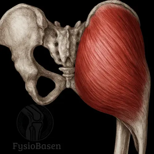
Origin
The gracilis has a broad origin involving several surfaces of the inferior pelvis:
Lower half of the body of the pubic bone
Entire surface of the inferior pubic ramus
Proximal part of the ischial ramus near the transition to the pubis¹
The muscle belly begins broad and flat and gradually converges into a thin, rounded tendon that continues distally.
Insertion
The course of the tendon gives the gracilis distinctive features at the knee. It passes:
Posterior to the sartorius
Anterior to the semitendinosus
Along the medial tibial epicondyle
The tendon inserts into the common tendinous structure pes anserinus together with the sartorius and semitendinosus. The insertion is located medially and proximally on the tibia, just below the joint line².
Topographical Relations
The gracilis lies in the medial thigh compartment and is immediately subcutaneous:
Lateral to the sartorius in its proximal part
Medial to the adductor longus and adductor magnus
Superficial to the pes anserinus bursa and the medial collateral ligament of the knee
The fascia lata covers the muscle, and the saphenous nerve and branches from the descending genicular artery pass through the fascial interval between the sartorius and gracilis⁴. Distally, the gracilis tendon may partially blend with the fascia of the medial head of the gastrocnemius.
Innervation
The gracilis is supplied by the obturator nerve via its anterior branch:
Nerve: Anterior branch of the obturator nerve
Spinal levels: L2–L4
Special note:
The obturator nerve also innervates the adductor longus and occasionally the adductor brevis. The gracilis receives several distinct nerve branches that follow different fibre bundles through the muscle, suggesting partial independent motor control of different muscle segments.
Blood Supply
The gracilis receives blood from multiple sources, providing good protection against ischaemia:
Primary source: Profunda femoris artery (via branches to the adductors)
Supplementary branches:
Medial circumflex femoral artery (proximal portion)
Femoral artery (distal portion)
The adductor artery enters the muscle on its lateral surface, approximately one third of the distance from the origin. This ensures a stable and evenly distributed blood supply, which is important for recovery and surgical procedures.
A Multi-Joint Muscle With Specialised Function
The gracilis is a rare muscle crossing both the hip and knee joints, giving it a unique role in movements requiring coordination between the pelvis and lower limb. It acts synergistically with the other adductors but is distinct in its direct influence on knee rotation and flexion.
Hip Joint: Adduction and Flexion
At the hip, the gracilis contributes to adduction, drawing the thigh toward the body’s midline. This is particularly important in activities requiring the legs to be pressed together, such as horseback riding, swimming, or running on sand or uneven terrain.The muscle also assists with slight hip flexion, although this function is weak compared with muscles such as the iliopsoas and rectus femoris.
Knee Joint: Flexion and Internal Rotation
At the knee, the gracilis has two clear roles:
Knee flexion, especially during the initial part of the swing phase in gait
Internal rotation of the tibia, which occurs only when the knee is flexed
This combination of movements is relevant in daily activities such as sitting down, crossing the legs, or adjusting body position during walking.
Stabilising and Postural Function
The gracilis also contributes to pelvic balance during gait, particularly when the contralateral limb is in the swing phase. With the foot planted, the gracilis works together with the hamstrings to rotate the femur and pelvis laterally, improving postural control and rotational stability.
During rapid changes of direction in sports such as football and handball, the gracilis is active in both acceleration and deceleration, helping stabilise the knee against internal rotation and resisting lateral displacement.
Contribution to the Pes Anserinus
The gracilis forms part of the pes anserinus tendon complex together with the sartorius and semitendinosus. This structure provides mechanical support to the medial aspect of the knee and contributes to controlled knee flexion and rotation. The pes anserinus also plays an important role in protecting against excessive valgus movement of the knee.
Injury-Prone Region
The gracilis is among the muscles most frequently involved in groin pain in athletes. Rapid and explosive movements may overload the muscle, particularly at its proximal tendon attachment to the pubis, resulting in adductor tendinopathy.It has also been observed that women generally have smaller gracilis muscle volume than men, which may influence the risk profile for knee injuries such as anterior cruciate ligament injury.
Activation and Strengthening of the Gracilis
The gracilis is a weak but functionally important muscle contributing to hip adduction, knee flexion, and medial rotation of the tibia. Because it crosses both the hip and knee joints, it must be trained in positions where both joints are engaged. Research shows that activation increases significantly when adduction is combined with knee flexion and internal rotation¹.
Adduction With Ball Between the Knees
Purpose: Basic activation of the gracilis through isometric contraction.
Execution:
Sit on a chair or lie supine with hips and knees at 90°.
Place a ball between the knees.
Squeeze the knees together for 5–10 seconds, relax, and repeat.
Interpretation: Easy to perform and provides safe activation without high load. Suitable early in rehabilitation.
Lateral Lunge With Resistance Band
Purpose: Dynamic strengthening of the gracilis through functional adduction.
Execution:
Stand with a resistance band around the ankles.
Take a long step to the side and lower into a lunge.
Push back in a controlled manner, focusing on activation of the medial thigh.
Interpretation: Increases gracilis recruitment through both eccentric and concentric work in movements where injury commonly occurs in sport.
Side-Lying Leg Raise With Foot Rotation
Purpose: Specific activation of the gracilis via combined hip adduction and internal knee rotation.
Execution:
Lie on the side with the lower leg extended.
Slightly rotate the foot inward and lift the leg straight upward.
Hold the top position for 2–3 seconds before lowering.
Interpretation: Electromyographic studies show greater gracilis activation with medial foot rotation compared with neutral or externally rotated positions¹.
Cable Adduction With Slight Knee Flexion
Purpose: Controlled strengthening with consistent resistance throughout the movement.
Execution:
Attach the cable at the ankle and stand in slight knee flexion.
Move the leg toward the midline in a slow, controlled motion.
Briefly hold the end position and return slowly.
Interpretation: Allows gradual progression of load. Slight knee flexion increases gracilis activation in addition to adductor longus and magnus².
Eccentric Training With Sliding Foot
Purpose: Prevention of strains and ruptures through eccentric strengthening of the gracilis.
Execution:
Stand with one foot on a slippery surface (towel or slide board).
Slide the leg out to the side while controlling the speed and engaging the medial thigh to brake the movement.
Push back to the starting position.
Interpretation: Eccentric training reduces the risk of adductor injuries and should be included in sports rehabilitation programmes³.
References
Standring, S. (2016). Gray’s Anatomy (41st ed.). Elsevier Churchill Livingstone.
Moore, K. L., Dalley, A. F., & Agur, A. M. R. (2014). Clinically Oriented Anatomy (7th ed.). Lippincott Williams & Wilkins.
Palastanga, N., & Soames, R. (2012). Anatomy and Human Movement: Structure and Function (6th ed.). Churchill Livingstone.
Nordin, J. S., Olsson, O., & Lunsjö, K. (2019). The gracilis tendon autograft is a safe choice for orthopedic reconstructive procedures. BMC Musculoskeletal Disorders, 20(1). doi:10.1186/s12891-019-2520-50(1). doi: 10.1186/s12891-019-2520-5