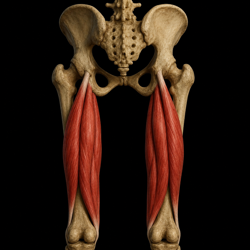
Iliacus
- Fysiobasen

- Jan 11
- 9 min read
Iliacus is the flat, triangular muscle that fills the inner surface of the pelvis and, together with psoas major, forms the powerful iliopsoas. This section provides a thorough overview of origin, insertion, anatomical relations, innervation, blood supply, basic function, clinical relevance, and the consequences of shortening.
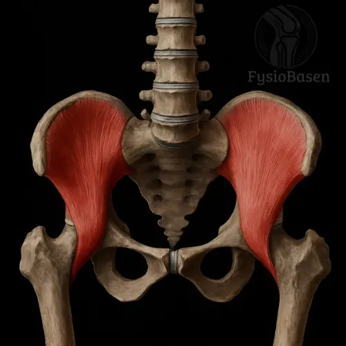
Origin
Iliacus has an extensive origin that contributes to its triangular shape and broad surface area:
The upper two thirds of the iliac fossa on the inner surface of the ilium
The inner lip of the iliac crest, near the anterior superior iliac spine
The lateral surface of the sacral body at the anterior sacroiliac ligament
An inferior fascicle from the iliolumbar ligament
Insertion
Distally, the fibers converge into a common tendon that passes deep beneath the inguinal ligament and blends with psoas major to form the common iliopsoas tendon. This tendon crosses the hip joint anteriorly and inserts on:
The lesser trochanter of the femur, often overlapping with fibers from psoas major
This location allows iliacus to transmit force efficiently from the pelvis to the femur during hip flexion.
Relations
The position of iliacus within the pelvis creates complex relationships with fasciae, nerves, and internal organs:
Anterior: Covered by the iliac fascia, separating the muscle from the peritoneum. The lateral femoral cutaneous nerve runs here, and the cecum (right) and descending colon (left) lie against the anterior surface of the muscle.
Medial: Along the inner border of the muscle runs the femoral nerve, closely adjacent to the lateral edge of psoas major. This junction forms the strong iliopsoas mass before it passes beneath the inguinal ligament.
Inferior: The fiber bundle passes beneath the inguinal ligament, curves deep past the subtendinous bursa, and blends with psoas major before tendon insertion.
Lateral: Bounded by fascial septa toward the inner surface of the pelvic brim, and channels for the iliolumbar artery and accompanying venous branches.
Posterior: Adjacent to the hip capsule and acetabulum, separated by a subtendinous bursa that protects against friction.
Innervation
Iliacus receives all of its nerve supply from the femoral nerve (L2–L4), which:
Passes beneath the inguinal ligament through the muscular lacuna
Gives off multiple motor branches to the deep, medial, and posterior surfaces of the muscle
Sends sensory branches to the hip joint capsule, important for proprioception
Injury or compression in this region results in weakened hip flexion and reduced sensation over the anterior thigh.
Blood Supply
The muscle has a rich arterial network that ensures oxygen delivery during both quiet standing and powerful movements:
Iliolumbar artery (from the internal iliac artery) provides the main supply via several branches along the surface of iliacus
Deep circumflex iliac artery supplies the upper portion and forms collaterals during hip flexion
Obturator artery sends small branches to the deepest muscle structures
Femoral artery contributes peripheral branches along the tendon toward the insertion on the lesser trochanter
This extensive vascularization supports good recovery but also carries a risk of large hematomas following trauma.
Function
Iliacus participates in multiple movements and stabilizing roles:
Hip flexion: With the trunk fixed, the muscle lifts the femur forward—essential for walking, running, stair climbing, and sit-ups.
Trunk flexion: With the femur fixed, iliacus pulls the trunk forward toward the thigh, contributing to forward bending in standing or supine positions.
Stabilization: During static standing, it helps control anterior pelvic tilt and counteracts excessive lumbar lordosis.
Force transmission: Together with psoas major, iliacus serves as a key link in transmitting forces between the trunk and lower limb, especially during explosive movements.
Movements Contributed to – Joints and Directions
The architecture of iliacus and its insertion on the lesser trochanter provide specific moment arms:
Hip flexion (sagittal plane): Primary movement; lifting the femur forward. Iliacus accounts for approximately half of total flexion force up to 90° of hip flexion; beyond this, psoas contributes relatively more.
Minimal external rotation (transverse plane): When the hip is deeply flexed (over ~70°), fiber orientation produces a slight external rotation. This is important in deep squats, martial arts kicks, and deep hip-opening positions.
Lateral stabilization (frontal plane): Although not a primary lateral mover, iliacus contributes—via the common tendon and connective tissue—to stabilizing the pelvis in the frontal plane during single-leg stance and gait, counteracting tendencies toward internal rotation.
Interaction With Other Muscles
Smooth and safe movement requires coordination between iliacus and several muscles:
Synergists in Flexion
Psoas major: Forms a common insertion and shares load evenly. During high-frequency movements (e.g., sprinting), they cooperate in rapid contraction and relaxation.
Rectus femoris: Assists when the knee is extended, such as in front kicks or high knee lifts.
Antagonists in Extension
Gluteus maximus and hamstrings: Eccentrically brake iliacus’ flexion moment and ensure controlled return of the leg.
Co-stabilizers
Transversus abdominis and erector spinae: Maintain a neutral spine during powerful hip movements, allowing iliacus to transmit force without pulling the pelvis into excessive tilt.
Gluteus medius/minimus: Act in the frontal plane to keep the pelvis level when iliacus is activated unilaterally.
When these muscles are imbalanced—due to weakness, overactivity, or injury—iliacus must compensate, potentially leading to excessive workload and overload.
Stabilizing vs. Mobilizing Function
Iliacus alternates between driving movement and locking the pelvis:
Mobilizing Role
In explosive activities (sprinting, jumping, kicking), rapid concentric contraction is required to lift the femur quickly. Iliacus’ long fibers and optimal moment arm for hip flexion at 30°–60° make it highly effective.
Stabilizing Role
In static exercises (plank with leg lift, single-leg squat), iliacus works isometrically to stabilize the pelvis, while its short, pennate fibers provide fine adjustments for small angular differences.
The ability to switch rapidly between these roles is crucial for both performance and injury prevention.
Movement Under Load and in Daily Activity
In daily life and sport, iliacus’ roles combine in a seamless chain:
Walking
Terminal swing (≈90° flexion): Iliacus eccentrically brakes femoral descent, damping impact.
Loading response (0–20°): Transition to concentric lifting to propel the body forward.
Stair Negotiation
Descending stairs: Requires marked eccentric control; iliacus acts as a damper as the body lowers.
Ascending stairs: Increases concentric force to lift the body, often combined with quadriceps activity.
Running and Sprinting
Swing phase: Rapid concentric contraction with quick cycles of contraction and relaxation.
Landing phase: Eccentric braking protects the hip and knee joints from shock.
Prolonged Sitting
Shortens iliacus through constant flexion, leading to tightness and reduced circulation.
Standing up requires reestablishing a full extension sequence to restore normal length–tension characteristics.
Further research indicates that optimal training distribution—alternating endurance and strength work, along with dynamic stretching—enhances iliacus’ capacity to meet these demands without overload.
Clinical Relationships
Problems involving iliacus often produce pain and functional limitations:
Tendinopathy and bursitis: Frequent high-load hip flexion without adequate recovery can irritate the iliopsoas tendon and inflame the subtendinous bursa, causing deep hip and groin pain worsened by passive stretch and knee extension.
Femoral nerve compression: Hemorrhage, hematoma, or muscle hypertrophy can compress the femoral nerve in the muscular lacuna, leading to quadriceps weakness, impaired knee extension, and reduced sensation over the anterior thigh.
Excessive tone and low back pain: Hypertonic iliacus pulls the pelvis anteriorly and increases lumbar lordosis, contributing over time to chronic low back pain.
Movement pattern disturbances: A shortened iliacus limits hip extension. Compensation via increased knee flexion or lateral hip motion may lead to knee and hip pain over time.
Shortening of Iliacus
With prolonged sitting, insufficient stretching, or unilateral sport without flexibility training, iliacus progressively shortens:
Increased anterior pelvic tilt: The pelvis rotates forward, lumbar lordosis increases, and low back pain develops.
Restricted hip extension: Evident during walking, running, and when lying flat on a bench with one leg hanging down.
Compensatory muscle activity: Hamstrings and gluteal muscles work harder to achieve full hip extension, leading to posterior thigh and gluteal pain.
Reduced daily function: Difficulty rising from a chair, stair climbing with extended knees, and instability during rapid direction changes.
Prevention and treatment involve systematic stretching with an eccentric focus, active awareness of pelvic position, and functional exercises such as pelvic lifts and dynamic hip extension. Part 2 will address detailed motor control, interaction with other muscles, and movement patterns under varying loads.
Clinical Findings and Typical Symptoms of Dysfunction
Iliacus dysfunction often presents with diffuse anterior hip and groin pain that may radiate toward the quadriceps. Patients frequently report stiffness and a “locked” sensation during hip flexion, particularly when attempting to lift the leg from supine or seated positions. Other common symptoms include:
Pain during resisted active hip flexion
Altered gait with shorter step length and increased hip flexion
Deep tenderness in the groin on palpation
Palpation, Examination, and Pain
Examination of iliacus requires precise localization of the iliac fossa:
Palpation Technique
The patient lies supine with hips and knees slightly flexed.
The therapist places fingertips deep into the iliac fossa, just medial to the anterior superior iliac spine.
The patient performs active hip flexion against light resistance; tenderness and pain quality on palpation indicate iliacus dysfunction.
Movement Testing
Passive hip extension stretch with the knee flexed to isolate iliacus
Thomas test to identify shortening and pain provocation during extension of the non-tested leg
Pain Patterns
Local pressure pain in the hip hollow
Sharp pain elicited during resisted hip flexion testing
Possible referred pain toward the medial thigh with irritation of the femoral nerve
Relevance for Sport, Rehabilitation, and Physiotherapy

Iliacus is critical in athletic movements that require explosive hip flexion and rapid step-up or stair-like actions. Injury or dysfunction may lead to substantial performance decline and increased injury risk.
Key sport-specific consequences:
Sprinting and running: Reduced hip flexion limits knee drive and stride frequency, increasing compensatory load on the hamstrings.
Jumping and power output: Insufficient iliacus force reduces vertical propulsion and shifts load toward the quadriceps and calf musculature.
Rehabilitation should follow a stepwise, load-progressive model:
Early phase: Pain modulation and gentle isometric activation (e.g., quadriceps setting with the hip in slight flexion).
Mid phase: Progressive eccentric loading (standing hip extension against resistance) combined with deep passive stretching into extension.
Late phase: High-velocity functional drills (high knees, fast step-ups) and sport-specific movement patterns.
Postural Function
In quiet standing, iliacus contributes to maintaining a mild anterior pelvic tilt, supporting lumbopelvic stability. When function is impaired, several maladaptive patterns may appear:
Overactivation of erector spinae, leading to increased lumbar lordosis and low-back pain
Flattening of the lumbar spine due to iliacus inhibition, increasing passive strain on posterior ligaments
Hip instability, predisposing individuals to knee hyperextension locking
Movement Patterns, Maladaptation, and Compensation
When iliacus does not function optimally, the body adapts through altered movement strategies:
Compensatory squat mechanics: Increased knee flexion to advance the limb, overloading quadriceps and knee joint
Increased lumbar rotation: Pelvic and trunk rotation substitute for limited hip flexion, contributing to chronic back symptoms
Asymmetric gait: Iliacus shortening reduces step length on the affected side, creating stride asymmetry
Prevention of maladaptive loading requires:
Restoration of normal iliacus length via eccentric-biased stretching
Neuromuscular retraining of hip and core musculature
Gradual exposure to functional loads across multiple planes
With systematic assessment and targeted rehabilitation, iliacus function can be restored, enabling pain-free activity with optimal movement control.
Exercises for Iliacus
1. Supine Pelvic Lift with Hip Flexion
Purpose: Activates iliacus and psoas major while emphasizing pelvic and lumbar control.
Execution:
Lie supine with knees flexed and feet flat.
Brace the core and slightly posteriorly tilt the pelvis.
Lift the pelvis a few centimeters.
Maintain pelvic height while extending one leg upward with the knee straight.
Lower with control.
Dosage: 8–12 repetitions per side, 2–3 sets.
Clinical effect: Improves coordination between pelvic stabilization and hip flexion while reducing compensatory dominance of rectus femoris.
2. Supine Straight Leg Raise
Purpose: Selective strengthening of iliacus during hip flexion with minimal quadriceps contribution.
Execution:
One leg flexed with foot on floor.
Opposite leg raised straight to ~45°.
Hold 1–2 seconds, lower slowly.
Dosage: 10–15 repetitions per side, 3 sets.
Clinical effect: Supports hip-flexor strength without excessive joint compression.
3. Hanging Leg Raise
Purpose: High-demand activation of iliacus with simultaneous core stabilization.
Execution:
Hang from a bar with stable trunk.
Raise legs to ~90° hip flexion.
Lower slowly without lumbar sway.
Dosage: 6–10 repetitions, 2–3 sets.
Clinical effect: High EMG activity in iliacus with strong neuromuscular control demands.
4. Standing Hip Flexion with Miniband Resistance
Purpose: Functional, weight-bearing activation of iliacus.
Execution:
Miniband anchored behind the body.
Lift leg forward to ~45° hip flexion while maintaining pelvic stability.
Dosage:12–15 repetitions per side, 3 sets.
Clinical effect: Enhances proprioception, balance, and hip-core coordination.
5. Lunge to High Knee Drive
Purpose: Integrates dynamic hip flexion with lower-limb strength and balance.
Execution:
Step into a forward lunge.
Drive upward into a high knee lift.
Alternate sides fluidly.
Dosage:10–12 repetitions per side, 3 sets.
Clinical effect: Simulates sport-specific movement chains and explosive hip flexion.
Progression Levels
Beginner: Supine drills, low-angle leg raises (~30°)
Intermediate: Band-resisted standing hip flexion, light ankle weights
Advanced: Hanging leg raises, lunge-to-knee-drive combinations
References
Cael, C. (2010). Functional Anatomy: Musculoskeletal Anatomy, Kinesiology, and Palpation for Manual Therapists.
Wolters Kluwer.Moore, K. L., Dalley, A. F., & Agur, A. M. R. (2014). Clinically Oriented Anatomy (7th ed.).
Lippincott Williams & Wilkins.Netter, F. (2019). Atlas of Human Anatomy (7th ed.).
Saunders.Palastanga, N., & Soames, R. (2012). Anatomy and Human Movement (6th ed.).
Churchill Livingstone.Standring, S. (2016). Gray’s Anatomy (41st ed.). Elsevier.