

Elbow Fracture – Olecranon Fracture

Olecranon fractures commonly occur as part of elbow injuries and are considered among the more straightforward intra-articular fractures to manage, both surgically and through physiotherapy. They are usually classified as intra-articular fractures, although extra-articular variants also occur.
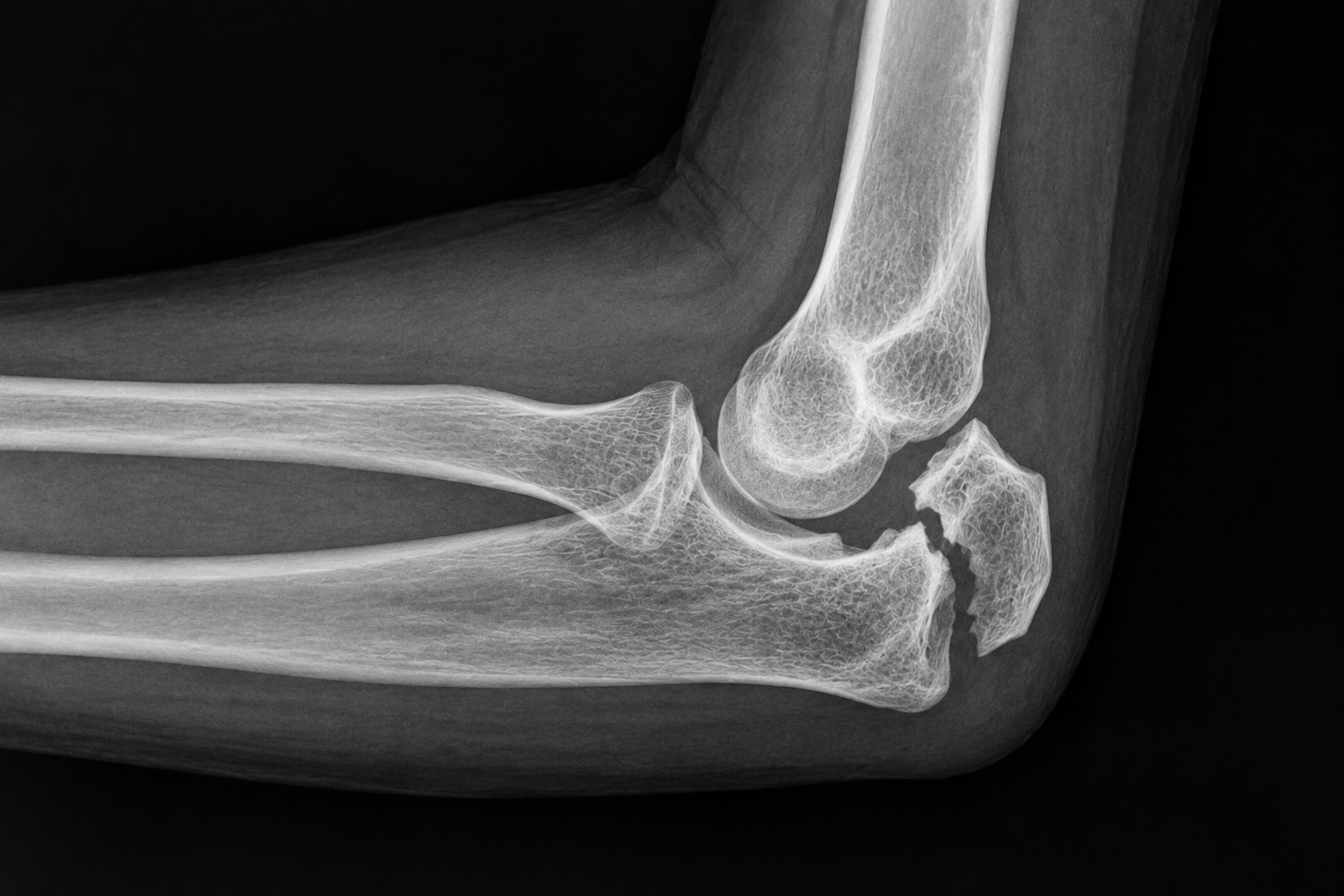
Classification and characteristics
Intra-articular fractures:
Most olecranon fractures are intra-articular.
Commonly associated with:
Joint effusion.
Hematoma in the injured area.
The injury may affect the stability and function of the elbow joint.
Extra-articular fractures:
Often include avulsion fractures.
Occur primarily in older patients, in whom osteoporosis may contribute to an increased risk of this type of injury.
Olecranon fractures are common after trauma or falls involving direct force to the elbow.
These fractures require careful assessment to ensure appropriate treatment because they may affect both joint structure and function.
Olecranon fractures are generally manageable, but individual assessment is required to optimize treatment and functional recovery.
Clinically relevant anatomy and epidemiology/etiology of olecranon fractures
Clinically relevant anatomy
The elbow consists of three separate joints, each with specific functions:
Ulnohumeral joint:
Function: Provides stability and enables flexion and extension of the elbow.
Radiocapitellar joint:
Function: Provides lateral stability and contributes to pronation and supination of the forearm.
Proximal radioulnar joint:
Function: Responsible for pronation and supination of the forearm.
Function of the olecranon
Triceps attachment:
The olecranon is an important insertion site for the triceps muscle, which plays a central role in the elbow extension mechanism.
Major articulation of the elbow joint:
The olecranon is a central component of elbow stability and function.
Epidemiology and etiology
Olecranon fractures result from direct or indirect mechanisms, and the mechanism may vary according to injury severity and the patient's activity level.
Mechanisms of injury
Direct injuries:
Fall onto the forearm:
The olecranon may be driven into the distal humerus when the elbow is hyperextended on an outstretched arm.
Penetrating injuries:
Injuries affecting the articular surface, for example penetrating or cutting trauma.
Indirect injuries:
Triceps contraction:
A forceful triceps contraction can cause an avulsion fracture when the olecranon fragment is pulled away together with the distal triceps attachment.
Sports injuries:
Athletes who generate high isometric forces through the arms, such as wrestlers and gymnasts, may be at increased risk.
This anatomical and epidemiological understanding provides important insight into how olecranon fractures occur and forms a basis for diagnosis and treatment.
Characteristics and clinical presentation of olecranon fractures
Olecranon fractures have specific clinical features that can help identify and classify the injury. A systematic approach to assessment and diagnosis is essential for appropriate treatment.
Clinical presentation
Main symptoms:
Patients with an olecranon fracture often have an inability to fully extend the arm.
Pain is usually localized over the elbow.
Observation:
Visible deformity around the elbow.
A shortened appearance of the forearm.
Swelling and bruising in the area.
Palpation:
Tenderness over the olecranon and proximal ulna.
A palpable defect or gap may be present at the fracture site.
Range of motion:
Limited elbow extension.
Classification: Mayo classification system
The Mayo classification assesses displacement, comminution and stability of the elbow joint. The system is divided into three types, each with two subgroups:
Type I:
Non-comminuted or minimally comminuted fractures.
A stable fracture without significant displacement.
Type II:
Displaced fracture with intact ulnar collateral ligaments.
Subdivided into:
Displaced versus nondisplaced.
Stable fracture.
Type III:
Fractures with disruption of the relationship between the forearm and humerus.
Unstable fractures because of injury to the medial collateral ligaments.
Subdivided into:
Displaced versus nondisplaced.
Diagnostic procedures
Physical examination:
Subjective interview:
Mechanism of injury: Direct blow to the elbow.
Observation:
Deformity, swelling and bruising around the elbow.
Palpation:
Tenderness and a possible gap at the fracture site.
Range of motion:
Limited elbow extension.
Special investigations:
X-rays:
AP and lateral views are standard for confirming the diagnosis and assessing fracture characteristics.
Olecranon fractures can be identified by their characteristic clinical features combined with systematic use of classification systems such as the Mayo classification, physical examination and imaging. This provides a strong basis for treatment planning and helps optimize prognosis.
Outcome measures and medical treatment of olecranon fractures
Outcome measures
Disabilities of the Arm, Shoulder and Hand (DASH):
Used to evaluate function and symptoms in the arm, shoulder and hand following treatment of olecranon fractures.
Provides insight into the patient's ability to perform daily activities and helps evaluate treatment effectiveness.
Medical treatment
Treatment of olecranon fractures depends on the degree of displacement, comminution and stability of the elbow joint. The primary goals are to restore elbow function, including the extensor mechanism, and reconstruct the articular surface to reduce the risk of osteoarthritis.
Type I fractures (non-comminuted and stable):
Treatment:
Immobilization for 2 weeks.
Symptomatic management, including pain relief and measures to reduce inflammation.
Risk:
Potential displacement as a result of traction from the triceps muscle.
Type II and III fractures (displaced and/or unstable):
Treatment:
Surgical fixation is required.
The goal is to provide stability, restore joint anatomy and function, and reduce complications such as osteoarthritis.
Prognostic factors
Primary elbow instability:
May indicate a risk of reduced elbow function and an increased likelihood of osteoarthritis.
Fracture morphology:
The complexity of the fracture influences treatment outcome and the rehabilitation process.
A structured approach to the management of olecranon fractures, based on classification and stability, is essential for achieving the best possible functional recovery and reducing long-term complications. The DASH score is a useful measure of treatment outcome and patient progress.
Surgical treatment of olecranon fractures
Surgical procedures for olecranon fractures are selected according to fracture complexity, patient needs and the degree of joint instability. The goal is to restore elbow function and stability.
Surgical methods
Tension band wiring (TBW):
Indication:
Simple, non-comminuted transverse fractures involving less than half of the olecranon articular surface.
Advantages:
Effective for stable fractures.
Complications:
Loss of fixation.
Nonunion or delayed union.
Infection, skin problems or prominent hardware.
Olecranon bursitis and radial-head subluxation.
Plate fixation:
Indication:
Comminuted fractures that require greater stability.
Advantages:
Good outcomes with respect to fracture union and a lower risk of implant-related complications.
Use:
Preferred for complex fractures that require increased mechanical stability.
Intramedullary screws:
Indication:
Simple, non-comminuted transverse fractures.
Advantages:
May provide improved pain, function and range of motion compared with tension band wiring.
Complications:
Reported less frequently than with TBW.
Olecranon resection and triceps advancement technique:
Indication:
The articular surface is too comminuted or osteoporotic for fixation, particularly in older or low-demand patients.
Method:
The triceps tendon is attached to the proximal edge of the remaining articular surface.
Prerequisites:
Intact structures such as:
Medial collateral ligament.
Interosseous membrane.
Distal radioulnar joint.
Possible complications
Surgical procedures may result in complications that affect patient function and prognosis:
Loss of extension:
Permanent loss of elbow extension may occur.
Heterotopic ossification:
Abnormal bone formation around the elbow joint.
Nonunion and malunion:
The fracture may fail to heal or may heal in an incorrect position.
Infection:
There is a risk of wound infection or deeper infection.
Surgical treatment of olecranon fractures requires an individualized approach based on fracture type and the patient's functional needs. Selecting an appropriate technique and managing complications correctly are important for achieving a good outcome.
Physiotherapy treatment after olecranon fracture
Careful postoperative follow-up is essential for optimal healing and functional recovery. The goals of physiotherapy are to restore daily activities and minimize functional limitations. Rehabilitation and bone healing usually take approximately 12 weeks.
Rehabilitation plan
Week 1: Inflammatory phase
Immobilization:
Splint or cast with the elbow at 60° of flexion to reduce skin tension.
Assessment:
Capillary refill, sensation, active and passive finger movement, and swelling.
Treatment:
Active movement of the shoulder, hand and fingers while the splint remains in place.
Edema management: Compression or elevation.
After 3–4 days:
Isometric wrist exercises.
Gentle active elbow flexion with the splint removed.
Active wrist movement.
Patient education:
Use the uninjured arm for self-care activities.
Week 2: Repair phase
Stability:
Elbow extension beyond 90° is not permitted while the arm is protected in a cast or splint.
Interventions:
The splint or cast is removed in most cases.
Gentle isometric exercises for the biceps.
Isotonic exercises for the fingers.
Weeks 4–6: Partially stable fracture
Active rehabilitation:
Active and active-assisted movement of the shoulder, elbow and wrist.
Avoid passive movements of the elbow.
Isometric exercises for elbow and wrist flexion and extension.
Activities of daily living:
Use the injured arm for light self-care and stabilization tasks.
Weeks 8–12: Clinical and radiographic healing
Physical progression:
Gradual weight-bearing through the injured arm.
Resistance exercises for the elbow flexors and extensors.
Active and active-assisted movement of the elbow and wrist in all planes.
Extension limitations:
Gentle passive stretching may be introduced if required.
A dynamic splint may be used to improve extension.
Clinical summary
Olecranon fractures are commonly caused by direct trauma or a forceful triceps contraction that may produce an avulsion fracture. Surgical treatment is often required, with specific physiotherapy guidelines used to support healing. Even with optimal rehabilitation, some patients may experience permanent loss of elbow extension, which is a recognized residual limitation after this type of fracture.
Sources:
Donegan RP, Bell J. Operative Techniques in Orthopaedics: Olecranon Fractures. Elsevier, 2010. p.17 -23.
Hoppenfeld S, Murthy VL. Treatment and Rehabilitation of Fractures. Lippincot, Williams & Wilkons, 2000.
Walters J, editor. Orthopaedics - A guide for practitioners. 4th Edition. Cape Town: University of Cape Town, 2010.
Meeusen R. Elleboogletsels in Sportrevalidatie. Kluwer, 2009. p.11-15,47.
Lavigne G, Baratz M. Fractures of the olecranon. Journal of the American Society for Surgery of the Hand, 2004;4(2):94-102.
Hutchinson DT, Horwitz DS, Ha G, Thomas CW, Bachus KN. Cyclic loading of olecranon fracture fixation constructs. Journal of Bone and Joint Surgery 2003;85(5):831-7.
Koslowsky TC, Mader K, Dargel J, Schadt R, Koebke J, Pennig D. Olecranon fracture fixation with a new implant: biomechanical and clinical considerations. Injury, 2009;40(6):618-24.
Kozin SH, Berglund LJ, Cooney WP, Morrey BF, An KN. Biomechanical analysis of tension band fixation for olecranon fracture treatment. Journal of Shoulder and Elbow Surgery, 1996;5(6):442-8.
Rommens PM, Küchle R, Schneider RU, Reuter M. Olecranon fractures in adults: factors influencing outcome. Injury, 2004;35(11):1149-57.


