Pectineus
- Fysiobasen

- Jan 11
- 7 min read
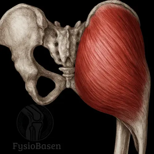
The pectineus is a flat, short, and broad muscle located at the transition between the anterior and medial compartments of the thigh. It is situated in the superomedial part of the anterior thigh, just inferior to the hip joint. Pectineus is a transitional muscle, both functionally and anatomically, and belongs to both the anterior and medial muscle compartments of the thigh. This is unusual and results in dual innervation and a distinctive anatomical course.
Pectineus lies medial to the iliopsoas muscle and lateral to the adductor longus muscle. The lateral border of the muscle is closely related to the medial circumference of the hip joint, while the medial border approaches the groin and the femoral fossa. Together with the iliopsoas muscle, pectineus forms the medial part of the floor of the femoral triangle (trigonum femorale).
The muscle has an almost square shape but may, in some individuals, be divided into two separate layers: a superficial (anterior) layer and a deeper (posterior) layer. These layers may have different innervation but lie closely together and function as a single muscle.
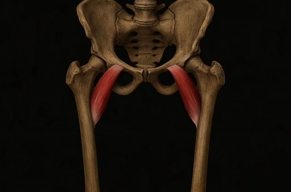
Origin
Pectineal line of the pubic bone (linea pectinea on the superior part of the superior pubic ramus)
Area immediately anterior to the pubic symphysis
The origin is broad and low, attaching along the anterior part of the pelvic ring, just lateral to the symphysis. The pectineus follows the superior border of the pubic bone and lies directly against the periosteum. The origin is often clearly visible during dissection and can be indirectly palpated through deep palpation medial to the groin.
Insertion
Pectineal line of the femur (linea pectinea on the posterior aspect of the proximal femur)
Proximal part of the linea aspera of the femur
The muscle inserts posteriorly on the femur along an oblique line that runs from the base of the lesser trochanter downward toward the linea aspera. This insertion line forms a continuous transition between the intertrochanteric line and the medial lip of the linea aspera. This anatomical relationship places pectineus close to the adductor group and explains why it is also classified as part of the medial compartment.
The insertion creates an oblique fiber orientation—lateral, inferior, and posterior—allowing the muscle to contribute to multiple hip joint movements depending on joint position.
Relations to Surrounding Structures
Pectineus has clinically and anatomically important relationships with surrounding structures:
Anteriorly: The deep fascia lata covers the muscle and separates it from the femoral artery, femoral vein, and great saphenous vein as they pass through the femoral triangle.
Laterally: It lies in close proximity to the iliopsoas muscle and the major vessels within the femoral canal.
Medially: The adductor longus muscle forms the other muscular component of the floor of the femoral triangle.
Posteriorly: It borders the adductor brevis, adductor magnus, and obturator externus muscles, as well as the anterior branch of the obturator nerve.
Pectineus is therefore strategically positioned at the intersection of several vascular and neural structures and is relevant both for functional control of the thigh and for understanding groin pain, impingement, and vascular compression.
The fascia lata encloses the pectineus and the other thigh muscles, and together with the intermuscular septa forms the internal boundaries between muscle compartments. Although pectineus belongs to both the anterior and medial thigh regions, the medial compartment lacks a clear fascial boundary and is instead defined by function and innervation.
Innervation
Pectineus is one of the few muscles in the body with dual innervation, reflecting its unique position within both the anterior and medial thigh compartments. This dual innervation contributes to its role in multiple movement directions and partly explains why clinical symptoms related to the pectineus are often diffuse or mixed.
Femoral nerve (L2–L3):This is the primary nerve supplying the superficial (anterior) part of the muscle. It belongs to the anterior compartment and originates from the lumbar plexus.
Obturator nerve (L2–L4) (in some individuals):This branch typically supplies the deeper (posterior) part of the pectineus. It arises from the lumbar plexus via the obturator canal and innervates the muscles of the medial compartment.
This combination of innervation allows pectineus to function both as a hip flexor (together with the iliopsoas) and as an adductor (together with the adductor muscles). This makes it particularly important during functional movements such as walking, rising from a chair, and stabilisation during single-leg stance.
Blood Supply
The blood supply to the pectineus is provided by two separate arterial systems, reflecting the muscle’s anatomical position and functional role:
Medial circumflex femoral artery:A branch of the femoral artery (or profunda femoris artery). It supplies the superficial part of the pectineus and courses laterally and posteriorly around the femoral neck, contributing to important anastomoses in the hip region.
Obturator artery (anterior branch):A branch of the internal iliac artery that passes through the obturator foramen. It supplies the deep portion of the muscle and also plays a role in the vascular network of the pelvis and adductor region.
Venous drainage generally follows the same pathways, with outflow via the femoral vein and obturator vein to the internal iliac vein.
In cases of rupture, strain, or trauma to the pectineus, these vessels may be affected, particularly in relation to bleeding associated with groin pain or hematoma formation in the proximal thigh.
Function
The pectineus has a complex and functionally versatile role in the hip joint. Its anatomical position and fiber orientation allow it to act not only in a single plane but to contribute to movements in both the sagittal and frontal planes, and to a certain extent in the transverse plane. This gives the muscle an important functional role in both dynamic movements and postural control.
Primary Movements
Hip Flexion
The pectineus works together with the iliopsoas and rectus femoris muscles to lift the thigh forward and upward at the hip joint. This occurs, among other situations, at the beginning of the swing phase during gait, when rising from a chair, and when lifting the leg to climb stairs.
Adduction
With increased hip flexion, the fiber orientation changes so that the muscle draws the femur medially toward the body’s midline. This occurs during crossing of the legs, bringing the legs together from an abducted position, and during single-leg balance in gait.
Secondary Movements
Internal and External Rotation
Studies suggest that the pectineus may contribute to both internal and external rotation of the hip joint, depending on the position of the leg. It is particularly debated whether internal rotation occurs in hip flexion and external rotation in a neutral position. This ambiguity is due to the oblique orientation of the muscle fibers relative to the femoral rotational axis.
Pelvic Stabilisation
As a postural muscle, the pectineus stabilises the pelvis in standing and during gait. This is particularly relevant during the phase of gait in which body weight is supported on one leg, where the muscles between the pelvis and femur maintain the hip joint centred within the acetabulum.
Clinical Relevance and Functional Significance
The pectineus is a transitional muscle, both anatomically and functionally. It connects the pelvis and femur and is integrated into complex movements requiring both force and control. During sports activities involving rapid changes of direction, kicking, or lateral movements, the muscle is subjected to significant load. It is therefore frequently involved in groin pain, particularly in cases of overuse, weakness, or muscular imbalance around the hip and pelvis.
In cases of excessive stretch, such as during unexpected lateral or forward leg movements, the muscle fibres of the pectineus may partially or completely rupture. Injury to the muscle typically presents as deep, medial groin pain with functional loss during hip flexion and adduction.
Research and Biomechanics
Biomechanical analyses and electromyographic studies show that the pectineus is activated during the initiation phase of gait and during stabilisation phases associated with rapid changes of direction. It demonstrates relatively high activation in functional exercises such as step-ups and supine straight-leg raises with slight abduction.
An EMG study from 2015 demonstrated significant pectineus activity during resisted hip flexion, particularly at angles between 30 and 60 degrees, confirming its role as an initiator of flexion. At the same time, MRI-based analyses have shown that the pectineus plays an important role in pelvic stabilisation in older adults and in patients with hip instability.
The muscle’s anatomical position and short fiber length also make it less susceptible to fatigue compared to larger muscle groups, and it functions as a “ligament-like” structure that contributes to joint stability under low-load conditions.
Targeted Exercises for the Pectineus
1. Side-Lying Hip Adduction
Purpose: Isolated activation of the pectineus and other hip adductors.
Execution: Lie on your side with the bottom leg extended straight and the top leg flexed over the other. Slowly lift the bottom leg off the floor and lower it in a controlled manner. Keep the pelvis stable.
Interpretation: Produces moderate to high EMG activation of the pectineus, particularly in the first 30 degrees of the lift¹.
Evidence: Studies show that this exercise produces higher isolated pectineus activity compared to standing cable or band adduction².
2. Standing Cable Adduction
Purpose: Functional strengthening of the pectineus with combined flexion and adduction.
Execution: Attach the cable at a low position and stand sideways to the machine. Move the leg diagonally forward and inward across the body while keeping the trunk stable.
Interpretation: The combination of adduction and flexion stimulates both primary functions of the pectineus. The movement simulates a phase transition during gait.
Evidence: EMG studies confirm higher activation when the movement occurs in the flexion plane³.
3. Sliding Hip Adduction Using a Friction Plate
Purpose: Dynamic control and strength of the adductor group, including the pectineus.
Execution: Stand with one foot on a friction plate. Slowly slide the leg out to the side while keeping body weight on the support leg, then draw the leg back in.
Interpretation: Produces eccentric loading during the outward slide and concentric loading during the return, effectively activating and strengthening the pectineus.
Evidence: Commonly used in rehabilitation of adductor injuries, with studies demonstrating good activation of both superficial and deep adductors⁴.
4. Active Hip Flexion With Ball Squeeze
Purpose: Activation of the pectineus during functional hip flexion combined with adduction.
Execution: Lie supine with a ball placed between the knees. Lift one leg into hip flexion while lightly squeezing the ball throughout the movement.
Interpretation: Isometric adduction combined with dynamic flexion produces high composite activation of the pectineus.
Evidence: Recommended in early rehabilitation of groin pain and adductor injuries⁵.
5. Step-Up With Medial Load Bias
Purpose: Strengthening of the pectineus in a functional, weight-bearing movement.
Execution: Perform step-ups while holding a weight (dumbbell) medially relative to the body. Actively guide the knee slightly inward during the movement.
Interpretation: Combining hip flexion and adduction in a weight-bearing position functionally activates the pectineus and synergistic muscles.
Evidence: Functional EMG studies show activation of the pectineus and its synergists during stair-climbing movements⁶.
References
Moore, K. L., Dalley, A. F., & Agur, A. M. R. (2014). Clinically Oriented Anatomy (7th ed.). Philadelphia, PA: Lippincott Williams & Wilkins.
Netter, F. (2014). Atlas of Human Anatomy (6th ed.). Philadelphia, PA: Saunders.
Palastanga, N., & Soames, R. (2012). Anatomy and Human Movement: Structure and Function (6th ed.). Edinburgh: Churchill Livingstone.
Standring, S. (2016). Gray’s Anatomy (41st ed.). Edinburgh: Elsevier Churchill Livingstone.