


Posterior Knee Pain

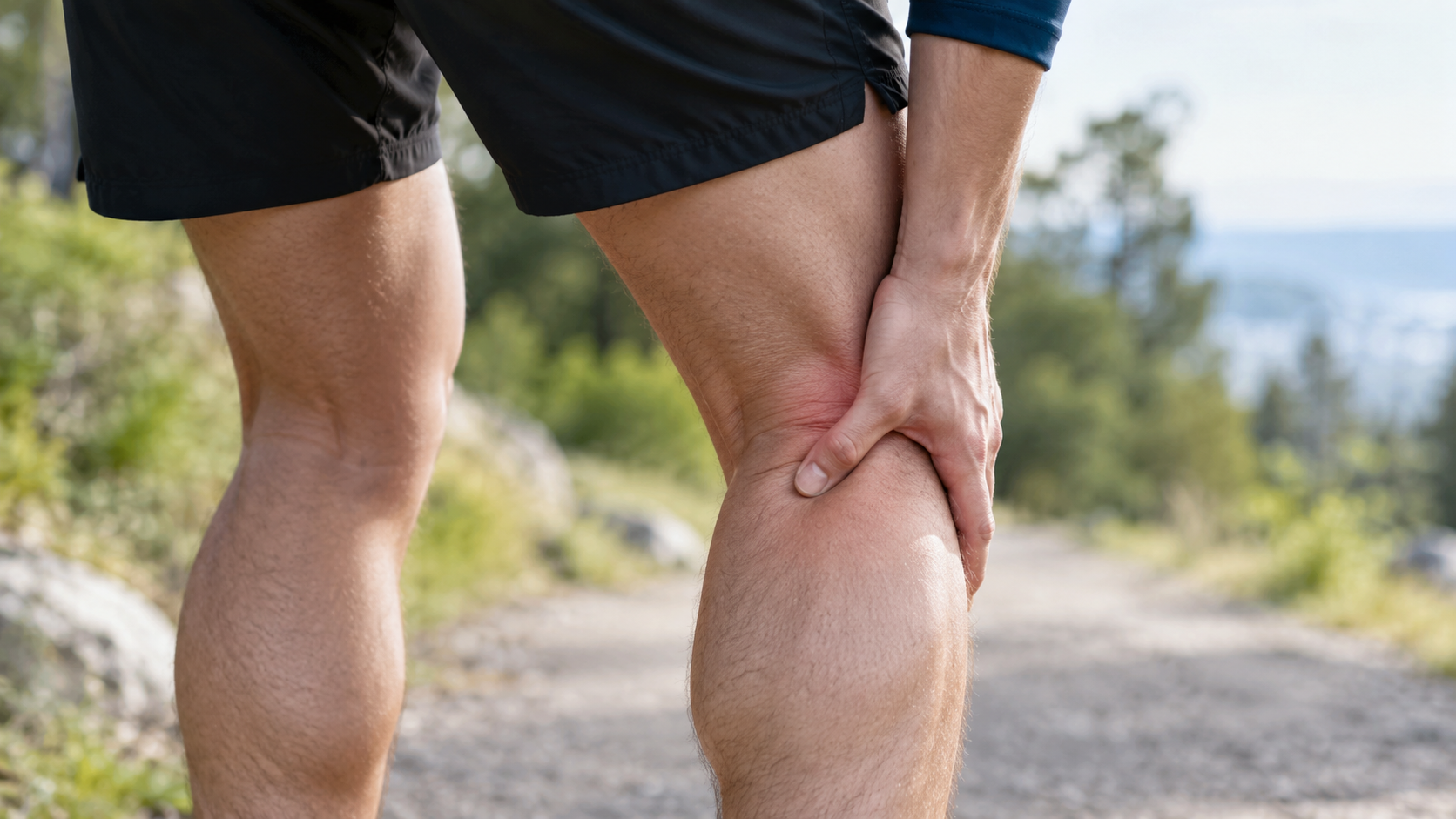
Posterior knee pain is a relatively common patient complaint, although pain in the anterior, medial, and lateral aspects of the knee is more frequent. The posterior knee contains a complex arrangement of muscles, ligaments, bursae, nerves, and vascular structures, so the causes of pain in this region can vary considerably. A thorough understanding of knee anatomy, combined with systematic clinical examination and a differential-diagnosis approach, is essential for identifying and managing the cause of posterior knee pain in a targeted manner¹.
Anatomically, the key active and passive stabilizers are located around and within the posterior aspect of the knee. Passive structures include the medial and lateral collateral ligaments (MCL and LCL), the anterior and posterior cruciate ligaments (ACL and PCL), and structures of the posterolateral corner. Active stabilizers include the hamstrings, popliteus, and gastrocnemius. The hamstrings consist of semimembranosus and semitendinosus medially and biceps femoris laterally. These muscles play a key role in knee flexion and dynamic stability. The popliteal region also contains important neurovascular structures, including the popliteal artery and vein, the small saphenous vein, and nerves such as the tibial and common peroneal nerves. Posterior knee pain may arise from injury or irritation of one or more of these structures.
Common causes of posterior knee pain
Muscular and tendinous injuries: Tendinopathies involving the hamstrings, gastrocnemius, and popliteus are among the most common causes of posterior knee pain. They are most often related to repetitive loading, particularly in sporting contexts. Semimembranosus tendinopathy typically causes a diffuse, aching posteromedial pain with tenderness on palpation below the joint line. Gastrocnemius injury produces pain with resisted knee flexion and during calf raises. The popliteus muscle, which plays an important role in “unlocking” the knee at the initiation of flexion, can be overloaded during activities requiring rotational control, especially downhill running.
Nerve injuries: Injury to the common peroneal nerve may occur with trauma to the posterolateral corner. This can cause pain, paresthesia, or reduced dorsiflexion strength. Neuropathic pain may also result from compression or entrapment of nerves in the popliteal fossa.
Ligament injuries: Injuries to the posterolateral corner and PCL can cause diffuse posterior knee pain, often associated with a sense of instability. These injuries commonly occur with hyperextension or rotational mechanisms.
Meniscal lesions: Injuries to the posterior horn of the meniscus, particularly the medial meniscus, may cause posterior knee pain, especially when the knee is extended after being flexed for a prolonged period. Such injuries often cause a sensation of catching or locking and may lead to localized swelling and reduced range of motion.
Bone-related conditions: Although less common, degenerative changes such as osteoarthritis or stress fractures of the tibia may cause pain behind the knee. Tumors such as osteochondroma, osteosarcoma, and synovial sarcoma should also be considered in persistent, unexplained pain, particularly in younger patients or when pain occurs at night and does not respond to conservative treatment.
Cysts and bursal involvement: The most common cause of swelling and pressure-related pain behind the knee is a Baker's cyst (popliteal cyst). This is a fluid-filled outpouching of the synovial membrane that often communicates with the knee joint and is secondary to intra-articular conditions such as osteoarthritis, meniscal lesions, or rheumatoid arthritis. The cyst may be palpable as a soft, rounded swelling and often causes pain during flexion as the bursa becomes compressed between the tibia and femur.
Vascular and neurological conditions: Popliteal artery entrapment syndrome, popliteal aneurysms, or deep vein thrombosis (DVT) can cause posterior knee pain, particularly when accompanied by swelling, erythema, or venous stasis. These conditions require prompt imaging and referral to a specialist.
Referred pain: Referred pain from the lumbar spine or hip may present as pain behind the knee. For example, L4 radiculopathy can cause posteromedial knee pain without local pathology. Patellofemoral disorders and patellar tendinopathy may also produce atypical radiation to the posterior knee.
Clinical examination and assessment
Palpation of the popliteal region may reveal swelling, tenderness, or asymmetry. A distinct, soft, fluctuant swelling may indicate a Baker's cyst. Tenderness over muscles and tendons points toward a musculoskeletal cause. Neurological examination of the lower limb should always be included, with assessment of sensation, strength, and reflexes, particularly when neurological or referred pain is suspected. If cystic lesions are suspected, ultrasound or MRI should be considered. In acute swelling and pain with redness and warmth, DVT must be excluded promptly.
For persistent symptoms, unclear clinical findings, or suspected tumor, MRI is the gold standard for further assessment. When there is obvious swelling behind the knee, ultrasound is a useful tool for identifying fluid collections or cysts.
Semimembranosus tendinopathy and hamstring strain
Semimembranosus tendinopathy (SMT) is a rare but important cause of chronic posteromedial knee pain, particularly in older patients or endurance athletes. The condition is often underdiagnosed, partly because of limited awareness and overlap with other disorders. SMT may develop primarily as an overuse injury during activities such as running and cycling, but it may also occur secondarily as a compensatory loading problem in the presence of underlying knee pathology, such as patellofemoral disorders¹.
Clinically, the condition is characterized by aching, localized pain on the posteromedial side of the knee, with clear tenderness on palpation just below the joint line. In the acute phase, pain may be intense and worsened by activities that load the hamstrings, including running, stair climbing, and sudden deep knee flexion³,⁴.
Hamstring strain is an acute injury that most commonly occurs during high-load sporting activities such as sprinting. It is characterized by sudden, sharp pain often described as a tearing sensation in the posterior thigh. Other symptoms include tightness, reduced strength, limited mobility, and painful walking.
Diagnostic assessment
The diagnosis of SMT is primarily clinical and based on a thorough history and palpation of the tendon insertion. When indicated, MRI can confirm the diagnosis and exclude other conditions. Outcome measures such as Lysholm Knee Score and Patient-Specific Functional Scale can be used to assess functional level and monitor progress during treatment.
In hamstring strain, clinical examination often provides a clear diagnosis. Patients typically present with localized swelling, tenderness on palpation, and reduced strength, particularly in knee flexion at 90°.
Medical treatment
For SMT, injection of 3 mL of 2% lidocaine and 10 mg triamcinolone at the tendon insertion may provide effective symptom relief³. Surgical intervention is rarely required, but tendon rerouting and reattachment may be considered in persistent cases.
For hamstring strain, the acute RICE principle is commonly used: rest, ice, compression, and elevation. NSAIDs may be used when needed for pain relief and reduction of inflammation. After the acute phase, heat may be used before exercise to increase tissue extensibility and circulation.
Physiotherapy treatment
SMT
Treatment of SMT is conservative in more than 90% of cases³. The first phase involves relative rest, cooling, NSAIDs, and pain-relieving measures. Exercise focuses on stretching and strengthening the hamstrings and surrounding musculature. Bodyweight exercise and eccentric strengthening are recommended.
Patients with genu valgum may benefit from a medial heel lift or motion-control shoes, for example Nike Zoom Structure Triax 15⁵,⁴. Such footwear provides additional support and can reduce overpronation, thereby decreasing mechanical stress on the tendon insertion.
Hamstring strain
Phase 1 (acute phase):
Submaximal isometric strengthening without pain.
Exercises are performed at 30°, 60°, and 90° of knee flexion.
Isometric knee flexion in a seated position.
The goal is normal gait and at least 50% strength compared with the unaffected side at 90° of flexion.
Phase 2 (rehabilitation):
Progression to eccentric strengthening and neuromuscular control.
Exercises include straight-leg deadlift, single-leg windmills and Nordic hamstring.
Nordic hamstring exercises can be performed with manual assistance or elastic support.
Phase 3 (late phase):
Focus on functional movement, plyometric training, and sport-specific activities.
Eccentric training in a lengthened position, for example using a cable, resistance band, or Biodex.
The patient should demonstrate full strength and pain-free performance of sport-specific movements before returning to sport.
Differential diagnoses for posterior knee pain:
Semimembranosus tendinopathy
Biceps femoris tendinopathy
Hamstring strain
Gastrocnemius strain
Popliteus strain or tendinitis
Posterior cruciate ligament injury
Posterolateral corner (PLC) injury
Baker's cyst
Knee bursitis
Common peroneal nerve injury
Popliteal artery entrapment syndrome or aneurysm
Deep vein thrombosis (DVT)
Referred pain from the hip, lumbar spine, sacroiliac joint, or radiculopathy¹
These conditions may present with overlapping symptoms, and careful clinical assessment is required to establish the correct diagnosis. It is particularly important to distinguish between intra-articular pathology, neurological and vascular causes, and muscular disorders because these may require very different treatment strategies.
Sources:
English S, Perret D. Posterior knee pain. Current reviews in musculoskeletal medicine. 2010 Oct 1;3(1-4):3-10.
Darlene Hertling, Randolph M. Kessler, “Management Of Common Musculoskeletal Disorders: Physical Therapy Principles and Methodes” (fourth edition), LippinCott Williams & Wilkins
Proper shoe fit: http://walking.about.com/od/prshoe/tp/shoemotion.htm
Proper shoe fit: http://www.runningshoesguru.com/2012/02/5-best-stability-running-shoes/
SANDRA J. SHULTZ, PEGGY A. HOUGLUM EN DAVID H. PERRIN. Examination of musculoskeletal injuries, third edition, Human Kinetics, 2010, 696 pages.
OHN C. GRIFFIN, Client-centered exercise prescription, Human Kinetics, 1998, 263 pages.
STEVEN J. FLECK, WILLIAM J. KRAEMER, Designing resistance-training programs, second edition, Human Kinetics, 1997, 275 pages.
DANIEL LORENZ, MICHAEL REIMAN, The role and implementation of eccentric training in athletic rehabilitation: tendinopathy, hamstring strains and ACL reconstruction, pubmed, march 2011, http://www.ncbi.nlm.nih.gov/pmc/articles/PMC3105370/, 27/11/2012,
BRANDON SCHMITT, TYLER TIM AND MALACHY MCHUGH, Hamstring injury rehabilitation and prevention of reinjury using lengthened state eccentric training: a new concept, pubmed, June 2012, http://www.ncbi.nlm.nih.gov/pmc/articles/PMC3362981/, 27/11/2012,


