

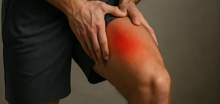
Quadriceps Contusion
- Fysiobasen

- Dec 24, 2025
- 4 min read
Quadriceps contusion is a common sports injury caused by a direct blow to the quadriceps muscle group, leading to significant soft-tissue damage. The impact results in rupture of muscle fibers within or near the injury site, often accompanied by hematoma formation, pain, and restricted motion. A contracted muscle absorbs impact energy better than a relaxed one, and therefore sustains less damage.
In sports without thigh and knee padding, such as football and rugby, quadriceps contusions represent a major cause of functional limitation. Although protective equipment can potentially reduce incidence, current research remains limited¹.
Two severe complications following quadriceps contusion include compartment syndrome and myositis ossificans²³.
Epidemiology
Quadriceps contusions occur about twice as often in men as in women.The highest incidence is seen in contact sports such as American football, soccer, and rugby, with most injuries occurring during competition rather than training.
Pathophysiology
The mechanism involves a direct blow to the quadriceps femoris, typically affecting the vastus intermedius muscle⁴.The pathophysiological process develops in a predictable sequence:
Myonecrosis and hematoma formation, followed by scar tissue development and later muscle regeneration.
Microscopic muscle fiber tears lead to bleeding and swelling within the anterior compartment⁵.
If larger hematomas are not treated properly, myositis ossificans may develop⁴.
Clinical Presentation
A thorough patient history revealing direct trauma, combined with a structured clinical examination, is essential for diagnosis.Typical findings include:
Skin discoloration and tenderness
Swelling and localized pain
Reduced range of motion
Difficulty bearing weight⁴
Classification
After muscle strain, contusions are the second most common quadriceps injury in sports.Severity is classified by knee flexion range 12–24 hours after trauma⁶⁸:
Grade | Active Knee Flexion | Gait | Description | Average Time to Return |
Mild | > 90° | Normal | Capillary rupture, mild tenderness, slight stiffness | ~6 days |
Moderate | 45–90° | Limping | Muscle bruising, swelling, pain, stiffness after rest | 5–6 days |
Severe | < 45° | Marked limp | Severe swelling, unable to walk without crutches, significant pain | > 60 days |
Physical Examination
Clinical assessment typically reveals:
Pain: Increasing over 24–48 hours, aggravated by active movement or knee flexion.
Observation: Limping gait pattern.
Palpation: Swelling, tenderness, discoloration, and possible palpable defect.
Circumference measurement: Compare muscle firmness and girth to the uninjured side.
Strength testing: Evaluate resisted knee extension and hip flexion.
Knee flexion: Key prognostic indicator⁴.
Provocation tests: Active straight-leg raise to assess extensor mechanism integrity.
Neurovascular status: Examine distal pulses and sensation to exclude compartment syndrome⁵¹⁰.
Outcome Measures
The Lower Extremity Functional Scale (LEFS) is widely used to assess function and recovery in lower-limb injuries.
Imaging
Advanced imaging assists in determining injury extent and complications:
MRI and Ultrasound: Evaluate soft-tissue trauma, hematoma, and edema¹⁰¹¹.
Ultrasound: Identifies localized hematoma and guides aspiration if needed.
X-ray: Useful to rule out bone injury or detect early myositis ossificans.
Risk Factors
Predisposing factors include:
Participation in contact or collision sports
Explosive or high-velocity movements
Inadequate warm-up or cool-down
Poor muscle flexibility or strength imbalances
Previous thigh, hip, or knee injuries
Bleeding disorders or use of anticoagulants¹²
Treatment
Acute Phase (0–48 hours)
Immediate care focuses on limiting bleeding and inflammation:
Immobilization with the knee in 120° flexion
Ice and compression for 24–48 hours⁵
Elevation and pain control
Avoid heat and massage initially
Short-term use of NSAIDs may reduce pain; prolonged use is discouraged.In severe injuries, early administration of NSAIDs has been shown to reduce the risk of myositis ossificans (evidence from hip arthroplasty studies).
If pain and restricted movement persist beyond 3–4 weeks, X-ray evaluation is recommended to exclude myositis ossificans¹¹¹³.
Physiotherapy Management
Rehabilitation is divided into three progressive phases:
Phase 1: Protection and Control
Compression bandage to limit hematoma
Immobilization at 120° knee flexion using wrap or brace
Gentle cryotherapy
Passive positioning to maintain flexibility and prevent stiffness
Phase 2: Early Activation
Begin gentle active movement after 24–48 hours
Introduce light stretching and isometric quadriceps contractions
Start active rehabilitation once 120° pain-free flexion is achieved
Phase 3: Functional Restoration
Gradual reintroduction of sport-specific training
Progressive resistance and dynamic strengthening
Focus on flexibility, coordination, and neuromuscular control
Return-to-Sport Criteria
Before clearance for full participation, the athlete should demonstrate:
Pain-free function
120° knee flexion with full hip extension
No residual swelling or weakness
Symmetrical performance on functional testing
Use of protective padding to prevent reinjury
Clinical Summary
Quadriceps contusions are common in contact sports and can lead to serious complications such as myositis ossificans or compartment syndrome if not managed correctly.Conservative treatment focusing on pain control, mobility restoration, and functional strengthening yields excellent outcomes in most cases.In severe or refractory cases, surgical intervention may be necessary.Physiotherapy plays a central role, emphasizing cryotherapy, soft-tissue management, and progressive functional training to ensure a safe return to sport.
Sources
Kary JM. Diagnosis and management of quadriceps strains and contusions. Current reviews in musculoskeletal medicine. 2010 Oct;3(1):26-31. : https://www.ncbi.nlm.nih.gov/pmc/articles/PMC2941577/
Kary JM. Diagnosis and management of quadriceps strains and contusions. Current reviews in musculoskeletal medicine. 2010 Oct 1;3(1-4):26-31
Christopher M. Larson, MD; Louis C. Almekinders, MD; Spero G. Karas, MD; William E. Garrett, MD, PhD. Evaluating and managing muscle contusions and myositis ossificans.2002 Feb;30(2):41-50.
Radiopedia Quadriceps Injury :https://radiopaedia.org/articles/quadriceps-injury
Orthobullets Quadriceps Contusion :https://www.orthobullets.com/knee-and-sports/3103/quadriceps-contusion
Faude O, Rößler R, Junge A. Football injuries in children and adolescent players: are there clues for prevention?. Sports medicine. 2013 Sep 1;43(9):819-37
G. Pasta, G. Nanni, [...], and S. Bianchi. Journal of ultrasound. Sonography of the quadriceps muscle: Examination technique, normal anatomy, and traumatic lesions. 2010 Jun; 13(2):76-84.
Huntoon EA. Essentials of Physical Medicine and Rehabilitation. InMayo Clinic Proceedings 2003 Apr 1 (Vol. 78, No. 4, p. 291). Elsevier.
Lee JC, Mitchell AW, Healy JC. Imaging of muscle injury in the elite athlete. The British journal of radiology. 2012 Aug;85(1016):1173-85.
Alonso A, Hekeik P, Adams R. Predicting a recovery time from the initial assessment of a quadriceps contusion injury. Aust J Physiother. 2000;46(3):167-77.
Shawn Bonsell,* MD, Paul T. Freudigman, MD, and Howard A. Moore, MD. Quadriceps Muscle Contusion Resulting in Osteomyelitis of the Femur in a High School Football Player. American journal of sports medicine. 2001;29(6)818-820.
Quadriceps Contusion (Cork Thigh).https://sma.org.au/resources-advice/injury-fact-sheets/quadriceps-contusion-cork-thigh/.
Diaz JA, Fischer DA, Rettig AC, Davis TJ, Shelbourne KD. Severe quadriceps muscle contusions in athletes: a report of three cases. The American Journal of Sports Medicine. 2003 Mar;31(2):289-93.


