Semitendinosus

Semitendinosus is a long, spindle-shaped muscle in the posteromedial thigh. It belongs to the hamstring group alongside the long head of biceps femoris and semimembranosus. These muscles share several features: they cross both the hip and knee, originate from the ischial tuberosity, and are supplied by the tibial division of the sciatic nerve. Semitendinosus is easily recognised by its long, prominent tendon, which gives the muscle its name.

The muscle contributes to hip extension, knee flexion and internal rotation, and also plays an important role in pelvic stability during standing and walking. Together with sartorius and gracilis, it forms the characteristic pes anserinus.
Origin
• Ischial tuberosity
Semitendinosus originates from the posterior and medial part of the ischial tuberosity, sharing a common tendon with the long head of biceps femoris. At this origin, semitendinosus usually lies medially and slightly deeper than biceps femoris.
The proximal third contains a fusiform muscle belly that gradually narrows into a long tendon around the middle of the thigh. The tendon travels distally and medially, behind the medial femoral condyle, and then passes behind the medial collateral ligament of the knee.
Insertion
• Medial surface of the proximal tibia through the pes anserinus
Distally, the tendon inserts on the upper medial surface of the tibia, just below the medial tibial condyle. Here it forms a shared tendon complex with gracilis and sartorius, called the pes anserinus—meaning ‘goose’s foot’ because of its characteristic fan-shaped arrangement.
Its position within the pes anserinus makes the tendon important for medial control and rotation of the lower leg at the knee, particularly in partial flexion. The tendon is separated from the medial collateral ligament by the bursa anserina, which is frequently involved in bursitis in this region.
Topographical relationships
In the thigh, semitendinosus lies medial to biceps femoris and superficial to semimembranosus. Together, these muscles form the superomedial boundary of the fossa poplitea. During knee flexion, semitendinosus is easily palpable as the most medial and posterior tendon among the three tendons on the medial side of the back of the knee.
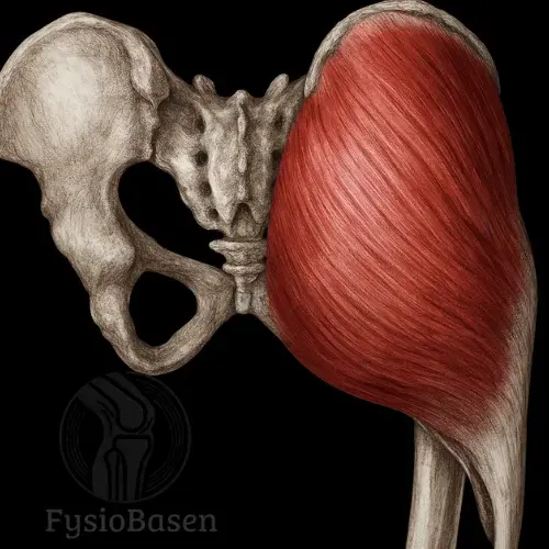
Proximally, the origin is covered by gluteus maximus, whereas the distal tendon lies relatively superficially. This makes it accessible for palpation and clinical testing and helps explain its frequent use as a graft in cruciate ligament reconstruction.
Relationship to the pes anserinus
Within the pes anserinus, semitendinosus lies:
– Deep and posterior to sartorius– Inferior and posterior to gracilis– Separated from the other tendons by individual bursae
These anatomical relationships are clinically important, particularly in pes anserine bursitis, in which inflammation beneath the tendon complex causes medial knee pain, often in runners or older adults with overuse. Tenderness over the tendon insertion and bursa is characteristic.
Innervation
Musculus semitendinosus is supplied by the nervus ischiadicus, specifically its tibial division (anterior roots, L5–S2). This division arises from the sacral plexus and follows the muscle through the posterior thigh compartment.
Motor innervation is delivered through deep branches entering the muscle belly directly. Function is controlled by both voluntary signals and reflex regulation during walking and balance adjustments. The sciatic nerve also supplies the long head of biceps femoris and semimembranosus, supporting coordinated hamstring function.
Sciatic nerve injury or compression, as in sciatica or lumbar disc herniation, may reduce semitendinosus strength and coordination. Clinically, this can cause reduced knee flexion and instability during hip extension, especially with heavy loading or rapid movement.
Blood supply
The muscle receives a combined blood supply from three main arterial sources:
1. Arteria profunda femorisThrough its first perforating artery, it supplies the proximal muscle. This branch is important for deep circulation to the posterior thigh muscles.
2. Arteria circumflexa femoris medialisProvides small but important contributions to semitendinosus and neighbouring muscles. Its anastomoses with gluteal and femoral branches are particularly relevant to regeneration and reconstructive surgery.
3. Arteria glutea inferior og arteria genus medialis inferiorThese provide distal contributions, particularly through branches of the arteria poplitea. Distal vessels supply the tendon complex around the pes anserinus, supporting healing after injury or surgical tendon harvest.
Compromised circulation, for example due to vascular disease or prolonged compression, may impair semitendinosus function, presenting as reduced endurance and longer recovery after activity.
Functional roles at the hip and knee
Semitendinosus has a dual functional role because it crosses both the hip and knee. The joint affected depends on which attachment is stabilised during movement.
With the lower leg fixed:– The hip is extended by pulling the femur backwards.– When the trunk is bent forwards, the muscle helps restore an upright position through hip extension.– It also acts as an internal rotator of the femur when the leg is slightly flexed.
With the pelvis fixed:– The knee is flexed as semitendinosus pulls the tibia backwards and upwards.– With the knee partly flexed, the muscle produces internal rotation of the lower leg. This is important for fine control during walking, rotation and balance adjustments.
Function depends on neuromuscular timing: the muscle is activated early in the swing phase of walking to slow hip flexion, and again at heel strike to stabilise the knee and promote hip extension.
Postural and dynamic stabilisation
In addition to producing movement, semitendinosus has an important postural role, particularly in standing. During single-leg stance, the muscle stabilises the pelvis in the frontal plane and resists lateral tilt, working with gluteus medius and other pelvic stabilisers.
During running and rapid direction changes, semitendinosus acts as a dynamic brake against anterior pelvic rotation and excessive hip extension. In this context, it supports the anterior cruciate ligament by resisting anterior translation of the tibia relative to the femur, particularly during weight-bearing with the knee partly flexed.
Clinical conditions and risk factors
Semitendinosus injuries often occur during explosive movements such as sprinting, jumping or sudden acceleration. The long tendon makes the muscle particularly susceptible to overstretching and rupture.
Common conditions include:
– Hamstring strain: common in sprinters and footballers, often at the musculotendinous junction.
– Tendon irritation at the pes anserinus: overuse, malalignment and poor biomechanics may cause chronic medial knee symptoms.
– Bursitis: inflammation of the anserine bursa can cause pain and swelling during stair use and squatting.
– Neurogenic hemiparesis: with unilateral weakness, for example after stroke, altered semitendinosus tone may affect gait.
The muscle’s anatomy and function also make it suitable as a tendon graft in cruciate ligament reconstruction. This can provide better postoperative knee function and less postoperative pain than a patellar tendon graft.
Palpation and clinical localisation
Semitendinosus is one of the hamstring muscles that can readily be palpated superficially. With the knee flexed against slight resistance, for example during an active squat, it appears as a distinct, tendinous structure on the medial side of the back of the knee.
It is best palpated:
– Proximally in the posterior thigh, medial to biceps femoris.– Distally as a taut tendon at the medial back of the knee, lateral to sartorius and gracilis.– At its pes anserinus insertion on the tibia, posterior to sartorius and inferoposterior to gracilis.
With overuse or injury, palpation may reveal local pain, firmness or tightness in the muscle or tendon. Pressure over the pes anserinus insertion often reproduces pain in bursitis or tendinopathy.
Assessment of strength and function
Manual muscle testing is often performed prone with the knee flexed approximately 90°. The patient resists while the therapist pushes the lower leg towards extension. Weakness presents as early fatigue or reduced resistance.
Neuromuscular control is best assessed during functional movements such as:
– Walking: look for dragging or asymmetrical hip and knee movement.
– Single-leg stance: reveals pelvic stability and postural control.
– Stair climbing: tests hip extension and knee flexion together under load.
If rupture, overstretching or neurological involvement is suspected, compare carefully with the unaffected side.
Special tests
1. Modified Thomas Test – flexibility assessment
The patient lies supine with one leg drawn towards the chest and the other hanging unsupported. If semitendinosus or other hamstrings are tight, the test hip will not drop into neutral extension and the knee will remain partly extended.
2. Puranen–Orava test – pain provocation through stretching
The patient stands with the hip flexed and the foot on a raised surface. Active forward bending with the knee straight may reproduce posterior thigh pain associated with tendinopathy or scar tissue.
3. Nordic Hamstring Test – eccentric control and rehabilitation
The patient kneels with the ankles held by a partner, then lowers the body forwards with control. The test requires considerable eccentric strength and is sensitive to weakness or residual symptoms after hamstring injury.
Clinical observations and malalignment
During gait analysis, semitendinosus weakness may produce:
– Reduced knee flexion during swing
– Compensatory hip flexion
– Pelvic tilt towards the opposite side during stance
Semitendinosus overcompensation, for example with weak gluteus maximus, may create an asymmetrical movement pattern and increase the risk of medial knee and hip overload.
If the muscle is shortened, the patient may show:
– Tight hamstrings in sitting or standing
– Reduced flexibility during passive testing
– Increased traction on the tibia and medial knee structures, particularly the MCL and pes anserinus
Postoperative follow-up and tendon grafting
Semitendinosus is frequently used as graft material for anterior cruciate ligament reconstruction. Harvesting rarely causes permanent functional loss, but postoperative management should consider:
– Gradual progression of eccentric strength
– Neuromuscular retraining of postural function
– Flexibility and tendon load tolerance
The early phase, 0–6 weeks, should focus on pain relief, swelling management and activation of neighbouring muscles, including the gluteals and quadriceps.
The intermediate phase, 6–12 weeks, includes gradual strengthening, weight-bearing exercise and increased attention to balance and control, whereas the later phase, after 12 weeks, includes sport-specific retraining and return to running, sprinting and changes of direction.
With appropriate progression and individual adaptation, most patients can resume full activity with good hip and knee function, even after tendon harvest.
Exercises for semitendinosus
Aim:
Semitendinosus is best activated by exercises involving hip extension and knee flexion, particularly during eccentric loading and with different hip and knee positions. This reflects its two-joint course and its postural and dynamic functions.
Research indicates that eccentric exercises, hip-extension exercises and positions involving long muscle lengths produce the highest semitendinosus EMG activity relative to the other hamstrings¹.
1. Nordic hamstring curl
Purpose: Strengthen semitendinosus and the other hamstrings eccentrically, particularly for injury prevention.
Technique: Kneel on the floor with the ankles secured by a partner or anchor. Keeping the body straight and knees on the floor, slowly lower the trunk forwards. Use the arms only as needed to arrest the fall.
Physiological effect: High semitendinosus activation during the eccentric phase. The muscle works in a lengthened position, supporting injury prevention and structural adaptation.
Research: The Nordic hamstring curl has documented effects on reducing hamstring injuries, particularly in sports involving acceleration and sprinting².
2. Glute-ham raise (GHR)
Purpose: Strengthen semitendinosus during both concentric and eccentric phases, with controlled hip and knee movement.
Technique: Using a GHR machine or modified bench, secure the lower legs, maintain a neutral hip position and lower forwards as in a Nordic curl, then return by activating the posterior chain.
Effect: Produces high semitendinosus activation, particularly near the bottom of the movement when both hip and knee are flexed.
Recommended for: Advanced trainees, patients in later rehabilitation and athletes with high posterior-chain force demands.
3. Romanian deadlift (RDL)
Purpose: Target semitendinosus and gluteus maximus through hip extension in a lengthened position.
Technique: Stand with the knees slightly bent, holding dumbbells or a barbell. Move the hips backwards while lowering the trunk, keeping the spine neutral. Return by driving the hips forwards.
Physiological focus: Strong activation throughout the hamstrings, with particularly effective loading at long muscle lengths for semitendinosus and semimembranosus.
Progression: Can be performed with a barbell, kettlebells or dumbbells, or as a single-leg variation.
4. Single-leg hip bridge with the foot on a bench
Purpose: Strengthen semitendinosus during functional hip extension while challenging stability and neuromuscular control.
Technique: Place one foot on a bench and raise the hips by pushing through the heel. Hold the other leg straight in the air.
Effect: Activates semitendinosus together with the gluteals and lumbar stabilisers. The control and postural activation requirements reflect its role during walking and running.
Variation: Use a resistance band for additional loading or suspension straps to introduce instability.
5. Swiss ball hamstring curl
Purpose: Train knee flexion and hip extension dynamically with control.
Technique: Lie supine with the heels on a large exercise ball. Raise the hips and roll the ball towards the buttocks by bending the knees. Return slowly.
Effect: Strong semitendinosus activation throughout the movement. Keeping the hips raised also maintains gluteal activation.
Rehabilitation: Suitable during early or intermediate rehabilitation after hamstring injury. Adjust loading through repetition count and ball size.
6. Standing cable kickback from a partial squat
Purpose: Specifically target semitendinosus by isolating hip extension in a lengthened position.
Technique: Attach an ankle strap to a cable and stand with your back towards the machine. Begin in a slight squat and press the leg backwards and upwards without substantially bending the knee. The movement should occur at the hip.
Effect: High semitendinosus activation, particularly with a low cable angle. Combining the movement with a slight squat further lengthens the muscle.
Tip: Maintain a slight forward trunk lean to keep tension in the muscle throughout.
Summary and progression
Semitendinosus training should include:
Eccentric exercises (Nordic, GHR)
Hip-extension exercises (RDL, kickbacks)
Functional movements (single-leg hip bridge, ball curl)
Postural control and loading at long muscle lengths
Combining these approaches supports activation, strength development and injury prevention. Select and adapt exercises according to the patient’s level, goals and function.
References:
Moore, K. L., Dalley, A. F., & Agur, A. M. R. (2014). Clinically Oriented Anatomy (7th ed.). Philadelphia, PA: Lippincott Williams & Wilkins.
Netter, F. (2019). Atlas of Human Anatomy (7th ed.). Philadelphia, PA: Saunders.
Palastanga, N., & Soames, R. (2012). Anatomy and human movement: structure and function (6th ed.). Edinburgh: Churchill Livingstone.
Standring, S. (2016). Gray's Anatomy (41st ed.). Edinburgh: Elsevier Churchill Livingstone.